


Moles Versus Melanoma: How to Tell the Difference
July 15, 2026
Moles Versus Melanoma: How to Tell the Difference

Moles are defined as benign skin growths formed by clusters of melanocytes, the pigment-producing cells in your skin. Melanoma is a malignant cancer that arises from those same cells and can spread to other organs if not caught early. Understanding the difference between moles and melanoma is not just reassuring. It is the first step toward protecting your life. The ABCDE criteria, developed by dermatologists as a clinical standard, give every person a practical framework for reading their own skin. Early detection dramatically improves survival outcomes, and that fact alone makes this knowledge worth carrying with you.
What are moles and why do they form?
Moles are clusters of melanocytes that grow together rather than spreading evenly across the skin. Most people develop between 10 and 40 moles over a lifetime, with the majority appearing before age 30. A typical benign mole is round or oval, smaller than 6 mm, evenly colored in shades of tan or brown, and has smooth, well-defined borders.

Not all moles look the same, and that variation is normal. Dermatologists classify moles into two broad types: common moles and atypical moles, also called dysplastic nevi. Common moles are stable, uniform, and rarely cause concern. Atypical moles are larger, have uneven color or irregular edges, and deserve closer attention. Atypical dysplastic nevi may be precursors to melanoma, but the vast majority never become cancerous.
The risk picture changes significantly with quantity. People with 10 or more atypical moles are 12 times more likely to develop melanoma than those without them. That statistic does not mean panic is warranted. It means those individuals need a consistent monitoring plan and regular dermatologist visits.
Moles can change slowly over years, and minor shifts in color or size are not always alarming. What matters is the pattern and pace of change. Sudden or rapid changes are the warning signal.
Key characteristics of a benign mole:
- Symmetrical shape, where one half mirrors the other
- Smooth, clearly defined borders
- Uniform color, typically tan, brown, or skin-toned
- Diameter smaller than 6 mm, roughly the size of a pencil eraser
- Stable appearance over months and years
Pro Tip: Photograph your moles monthly in the same lighting and position. Side-by-side comparison over time reveals changes that are invisible to memory alone.
How does melanoma differ from a benign mole?
Melanoma is malignant melanocyte proliferation. Unlike a mole, which stays contained, melanoma cells invade surrounding tissue and can travel through the lymphatic system or bloodstream to reach distant organs. That capacity to metastasize is what makes melanoma the most dangerous of all skin cancer types.
One of the most important facts about melanoma is where it starts. 70% to 80% of melanomas arise as entirely new spots on the skin, not from existing moles. That finding overturns the common assumption that you only need to watch your current moles. Any new pigmented growth deserves attention, not just the ones you have had for years.
Melanoma presents in several forms, and appearances vary by type:
- Superficial spreading melanoma is the most common type. It grows outward across the skin surface before going deeper, often showing multiple shades of brown, black, pink, or red.
- Lentigo maligna melanoma typically appears on sun-damaged skin in older adults as a flat, slowly expanding patch with uneven color.
- Nodular melanoma grows downward quickly and often appears as a raised, dark, or even skin-colored bump. It is the most aggressive form.
- Acral lentiginous melanoma develops on the palms, soles, or under nails and is more common in people with darker skin tones.
Biologically, melanoma cells lose the normal controls that stop growth. They divide without limit and produce enzymes that break down the tissue barriers that would otherwise contain them. This is why a melanoma caught at stage I has a very different prognosis than one caught at stage III or IV.
A note on pain: Painful moles are usually caused by non-cancerous issues like friction or infection. However, persistent pain lasting more than a week in any pigmented spot is a reason to see a dermatologist, not to wait.
What is the ABCDE criteria and how do you use it?
The ABCDE criteria are the standard clinical tool for identifying potential melanoma during a skin check. Dermatologists developed this framework so that both clinicians and the general public can apply a consistent, evidence-based method to evaluate pigmented spots.
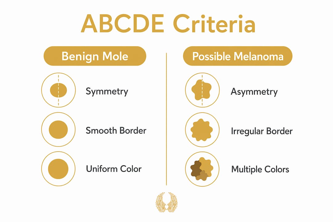
| Letter | What it stands for | What to look for |
|---|---|---|
| A | Asymmetry | One half does not match the other |
| B | Border | Edges are ragged, notched, or blurred |
| C | Color | Multiple shades of brown, black, red, white, or blue |
| D | Diameter | Larger than 6 mm, about the size of a pencil eraser |
| E | Evolution | Any change in size, shape, color, or new symptoms |
Each letter captures a distinct warning signal. Asymmetry matters because benign moles grow evenly. Irregular borders suggest uncontrolled cell growth at the edges. Multiple colors indicate different depths and rates of melanocyte activity. Diameter above 6 mm is a threshold, not a guarantee, since some melanomas are smaller. Evolution is arguably the most important criterion because change over time is the clearest sign that something is wrong.
The ABCDE framework has real limitations. Some melanomas, particularly nodular melanoma, do not fit the classic pattern. They may be small, symmetrical, and uniformly dark. This is why dermatologists also use the “ugly duckling” sign as a complementary tool. The ugly duckling sign identifies moles that look noticeably different from all the others on your body. If one spot stands out in color, size, or shape compared to your other moles, that outlier deserves professional evaluation.
Pro Tip: Do not wait for a spot to check every ABCDE box before seeing a doctor. One clear warning sign is enough reason to make an appointment.
How and when should you monitor your moles?
Monthly skin self-exams are the foundation of early melanoma detection. A self-exam takes about 10 minutes and requires good lighting, a full-length mirror, a hand mirror for hard-to-see areas, and a consistent routine. Check your entire body, including the scalp, between toes, under nails, and the back of your legs.
Signs that require a dermatologist visit:
- Any new mole appearing after age 30
- A mole that changes in size, shape, or color over weeks or months
- A spot that bleeds, itches, or crusts without an obvious cause
- A growth that looks different from all your other moles
- Any pigmented spot that does not heal within a few weeks
New moles appearing in adults over 30 are a specific warning sign because mole development naturally slows after that age. A new growth later in life has a higher chance of being something other than a routine mole. This does not mean every new spot is cancer. It means every new spot after 30 deserves a professional look.
When you visit a dermatologist, they will often use dermoscopy, a technique that uses a handheld magnifying device with polarized light to see structures beneath the skin surface. Dermoscopy improves mole visualization significantly but cannot replace biopsy for a definitive diagnosis. If a spot looks suspicious under dermoscopy, the next step is a biopsy.
A biopsy removes a small sample of tissue for laboratory analysis. Visual assessment alone cannot confirm melanoma. Biopsy is the only method that delivers a confirmed diagnosis. Biopsies also evaluate tissue depth and staging, which directly guide treatment decisions and prognosis.
“Many biopsied pigmented growths are benign. A biopsy is a precautionary diagnostic step, not automatic confirmation of cancer.” — Merck Manual Consumer
Key Takeaways
Distinguishing moles from melanoma requires knowing the ABCDE criteria, monitoring your skin monthly, and seeking professional evaluation for any spot that changes, stands out, or appears new after age 30.
| Point | Details |
|---|---|
| ABCDE criteria | Use Asymmetry, Border, Color, Diameter, and Evolution to evaluate every pigmented spot. |
| Most melanomas are new spots | 70–80% of melanomas arise as new growths, not from existing moles. |
| Atypical moles raise risk | Having 10 or more atypical moles increases melanoma risk 12 times. |
| Biopsy confirms diagnosis | Visual checks and dermoscopy guide suspicion, but only biopsy confirms melanoma. |
| New moles after 30 need evaluation | Mole development slows after age 30, so new growths later in life deserve prompt attention. |
What we have learned from watching people watch their skin
At the Hippocratic Cancer Research Foundation, we have seen something quietly powerful happen when people learn to read their own skin. They stop feeling helpless. They start showing up to appointments earlier. And earlier appointments save lives.
The most common misunderstanding we encounter is the belief that melanoma always looks dramatic. People expect something obviously alarming. The truth is that early-stage melanoma can look like a slightly irregular freckle. That is exactly why the ABCDE criteria and the ugly duckling sign exist. They give you a language for what your eyes are already noticing.
We also want to say this plainly: a biopsy is not a verdict. We have watched people delay getting a suspicious spot checked because they feared what the answer might be. That fear is understandable. But a biopsy is information, and information is what gives doctors the ability to act. The earlier that information arrives, the more options exist.
The advances happening right now in melanoma treatment at institutions like the Robert H. Lurie Comprehensive Cancer Center of Northwestern University are genuinely hopeful. Immunotherapy and targeted therapies have changed the prognosis for many patients. But those treatments work best when the disease is caught before it spreads. Self-awareness combined with expert consultation is not just good advice. It is the most powerful tool we have.
Trust what you see. Act on what concerns you. We are here, and we believe in the power of early action.
— HCRF
The HCRF’s mission in melanoma awareness and research
The Hippocratic Cancer Research Foundation supports bold, unconventional cancer research at the Robert H. Lurie Comprehensive Cancer Center of Northwestern University. Our mission is to fund the kind of work that changes outcomes, and melanoma research is at the heart of that effort.

Public awareness is one of the most powerful forces in early detection. When people understand what to look for, they seek help sooner. When they seek help sooner, survival rates rise. We believe that education and research are two sides of the same coin. Visit the Hippocratic Cancer Research Foundation to learn more about our work, find resources on skin cancer awareness, and join a community united by hope and determination. Together, we carry the light of that hope forward.
FAQ
What is the main difference between a mole and melanoma?
A mole is a benign cluster of melanocytes that stays contained, while melanoma is a malignant cancer that can invade tissue and spread to other organs. The key visual differences follow the ABCDE criteria: asymmetry, irregular borders, multiple colors, large diameter, and evolution over time.
Can melanoma develop on skin with no existing moles?
Yes. Between 70% and 80% of melanomas arise as entirely new spots rather than from pre-existing moles. Any new pigmented growth on your skin deserves attention, regardless of your mole history.
How do I know when to see a dermatologist about a mole?
See a dermatologist if a mole changes in size, shape, or color, bleeds or itches without cause, or looks noticeably different from your other moles. Any new mole appearing after age 30 also warrants professional evaluation.
Does a biopsy mean I have cancer?
No. A biopsy is a diagnostic step, not a diagnosis. Many biopsied pigmented growths turn out to be benign. The biopsy simply gives your doctor the tissue information needed to confirm or rule out melanoma.
What is the ugly duckling sign?
The ugly duckling sign refers to a mole that looks noticeably different from all the other moles on your body. That outlier, regardless of whether it meets all ABCDE criteria, deserves a professional evaluation because it stands apart from your personal baseline.
Recommended

Discover how corporate matching gifts can double your donations to nonprofits like the Hippocratic Cancer Research Foundation. Check your eligibility today!
Read More →
Practical guidance for researchers on precision medicine trials: prioritize the screening funnel, FDA master protocol rules, sequencing speed, and...
Read More →