


Does Tumor Size Determine Cancer Stage? What You Need to Know
July 25, 2026
Does Tumor Size Determine Cancer Stage? What You Need to Know

Tumor size partially determines cancer stage, but it is never the whole story. The TNM staging system evaluates three distinct factors: the primary tumor’s size and extent (T), lymph node involvement (N), and distant metastasis (M). A tumor can be quite large and still be classified as Stage I or Stage II if it has not spread to lymph nodes or distant organs. Conversely, a small tumor that has already seeded distant sites is Stage IV, regardless of its size.
Here is what that means in practice for you or someone you love:
- Tumor size matters but is one component of a three-part equation.
- Lymph node involvement can push a cancer to a higher stage even when the primary tumor is small.
- Distant metastasis automatically places a cancer at Stage IV, no matter the tumor’s dimensions.
- Large does not equal late-stage. A breast tumor with no spread may still be Stage II.
- Stage and grade are different things. Grade describes how abnormal the cancer cells look; stage describes how far the disease has traveled.
Understanding this distinction is not just reassuring. It is the foundation for making sense of a diagnosis, asking the right questions, and knowing what comes next.
Table of Contents
- What cancer staging actually means for your care
- How the TNM system measures tumor size and extent
- How tumor size relates to stage and what it means for prognosis
- Why lymph nodes and metastasis matter as much as tumor size
- Stage vs. grade: two different ways of understanding cancer
- Tests your doctor uses to measure tumor size and assign a stage
- What recent research tells us about tumor size and staging
- Support the research that changes what staging can do
- Key Takeaways
What cancer staging actually means for your care
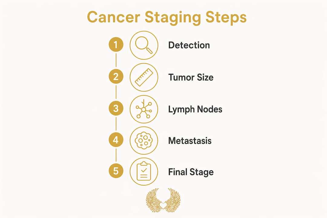
Cancer staging is the process of determining how far a cancer has grown and spread within the body. Oncologists assign a stage at diagnosis to answer one essential question: Where is this cancer, and how much of the body does it involve?
That answer shapes everything. It guides which treatments are appropriate, whether surgery is feasible, and how aggressively a medical team needs to respond. Staging also creates a shared language across hospitals, research institutions, and clinical trials, so that a “Stage II breast cancer” in Chicago means exactly the same thing as it does in Houston.
Staging is assigned at diagnosis and remains fixed for research consistency, even if the disease progresses later. That permanence can feel counterintuitive, but it allows researchers to compare outcomes across thousands of patients over decades. Without that consistency, the data that drives better treatments would collapse into noise.
One more thing worth knowing: staging systems are not universal. They are standardized within cancer types, but the thresholds and criteria differ between, say, lung cancer and colorectal cancer, because each disease behaves differently in the body.
How the TNM system measures tumor size and extent
The TNM classification is the most widely used cancer staging framework in the world. Each letter represents a separate dimension of the disease.
- T (Tumor): The size and local extent of the primary tumor, rated T0 through T4.
- N (Nodes): Whether cancer has reached nearby lymph nodes, rated N0 through N3.
- M (Metastasis): Whether cancer has spread to distant organs, rated M0 (no spread) or M1 (spread present).
The T category is where tumor size most directly enters the picture. T0 means no evidence of a primary tumor. T1 through T4 reflect increasing size or invasion into surrounding tissue, with T4 representing the most locally advanced disease. TX is recorded when the tumor cannot be measured.
Pro Tip: Physicians select the most accurate tumor size from imaging or pathology rather than simply the largest measurement available, specifically to avoid overstaging a patient.

Size thresholds vary by cancer type, which is one reason the system can feel complex. Here is how T categories map across two common cancers:
| T Category | Breast Cancer | Non-Small Cell Lung Cancer |
|---|---|---|
| T1 | Small | Small |
| T2 | Medium | Medium |
| T3 | Large | Large |
| T4 | Any size with chest wall/skin involvement | Any size invading mediastinum, heart, or great vessels |
These T category definitions vary by cancer type, which is why a 4 cm tumor might be T2 in one cancer and T3 in another. The biology of each disease determines where the meaningful thresholds fall.
Clinical staging (before surgery) and pathological staging (after surgical removal and lab analysis) can differ. Imaging sometimes overestimates tumor size, and the discrepancy between clinical and pathological staging is common and expected. Both provide valuable information, and together they give the most complete picture.
How tumor size relates to stage and what it means for prognosis
Smaller tumors tend to correlate with earlier stages, and earlier stages generally carry better prognoses. That relationship is real and well-documented, but it is not a simple one-to-one rule.
Research on non-small cell lung cancer illustrates this clearly. A study published in BMC Cancer found that 70.79% of tumors ≤20 mm were classified as N0M0 (no lymph node involvement, no metastasis), compared to only 33.33% of tumors measuring 71 mm or larger. Smaller tumors were far more likely to be caught at an early, localized stage. But “more likely” is not “always.”
What this means for prognosis:
- Stage is the stronger predictor of survival outcomes than tumor size alone.
- A large tumor with no lymph node spread (T3 N0 M0) may still be surgically curable.
- A small tumor that has reached distant organs (T1 N0 M1) is Stage IV with a very different outlook.
- Prognosis also depends on cancer type, grade, molecular markers, and the patient’s overall health.
The takeaway is this: tumor size influences stage, and stage influences prognosis, but size by itself is not a reliable severity marker. Two patients with the same tumor diameter can face entirely different treatment paths depending on what the lymph nodes and imaging reveal.
Why lymph nodes and metastasis matter as much as tumor size
Lymph node involvement and distant metastasis are the factors that most dramatically shift a cancer’s stage, and they can do so independent of how large the primary tumor is.

The N component of TNM captures whether cancer cells have traveled from the primary tumor into nearby lymph nodes. N0 means no node involvement. N1, N2, and N3 reflect increasing numbers of affected nodes or spread to more distant nodal regions. Even a relatively small primary tumor classified as T1 can push a cancer to Stage III if multiple lymph nodes are involved.
The M component is the most decisive of all. Stage IV is defined by distant metastasis, meaning cancer has spread to organs or tissues far from the original site, regardless of the primary tumor’s size. A tumor that has seeded distant organs is Stage IV. A large tumor confined to the breast with no nodal spread is not.
Why does this matter so much for treatment?
- Lymph node involvement often determines whether radiation to regional nodes is added after surgery.
- Metastatic disease typically shifts treatment from curative intent to disease management.
- The number and location of affected lymph nodes can influence the extent of surgical resection.
- Distant metastasis opens the door to systemic therapies: chemotherapy, targeted therapy, or immunotherapy.
Size alone is insufficient to make any of these decisions. The full TNM picture is what drives the clinical conversation.
Stage vs. grade: two different ways of understanding cancer
Stage and grade are both used to describe cancer, but they answer completely different questions. Confusing them is one of the most common misunderstandings patients carry into their first oncology appointment.
Stage describes the anatomical extent of the disease: how large the tumor is, whether it has invaded nearby tissue, whether lymph nodes are involved, and whether it has spread to distant sites. It is a map of where the cancer is in the body.
Grade describes the cellular appearance of the cancer. Pathologists examine cancer cells under a microscope and assess how abnormal they look compared to healthy cells. Low-grade tumors have relatively normal-appearing cells and are considered well-differentiated. High-grade tumors have markedly abnormal cells, are poorly differentiated, and tend to grow and spread more aggressively.
A cancer can be low-stage but high-grade, which means it has not spread far yet but its cells are aggressive and likely to do so. The reverse is also possible. Clinicians use stage to direct treatment intensity and grade to understand how the cancer is likely to behave over time. Both pieces of information shape the treatment plan, but they are not interchangeable.
Tests your doctor uses to measure tumor size and assign a stage
Determining tumor size and cancer stage requires multiple types of information gathered through different methods. No single test tells the whole story.
Imaging is usually the starting point:
- CT scan (computed tomography): Provides detailed cross-sectional images of tumors and nearby structures.
- MRI (magnetic resonance imaging): Particularly useful for soft tissue detail and brain or spinal involvement.
- PET scan (positron emission tomography): Detects metabolically active cancer cells, helping identify distant spread.
- Ultrasound: Often used for superficial tumors or to guide biopsies.
Beyond imaging, tests to determine stage include:
- Biopsy: Tissue sampling to confirm cancer type, grade, and molecular markers.
- Surgical exploration: Sometimes the only way to fully assess extent, particularly in abdominal cancers.
- Physical examination: Can detect palpable lymph nodes or masses, though it has clear limitations in accuracy.
- Blood and tumor marker tests: Support staging in certain cancers, such as PSA in prostate cancer or CA-125 in ovarian cancer.
Pathological staging, performed after surgical removal of the tumor, is generally more accurate than clinical staging done before surgery. Imaging can overestimate tumor size due to surrounding inflammation or edema, and accurate measurement is critical because even a few millimeters can shift a tumor from one T category to another.
What recent research tells us about tumor size and staging
The science on tumor size and staging has grown considerably more nuanced over the past two decades. Early assumptions that bigger always meant worse have given way to a more layered understanding.
The non-small cell lung cancer study referenced earlier is one of the clearest demonstrations of the size-stage relationship in the literature. The sharp drop from 70.79% N0M0 status in small tumors to 33.33% in larger ones confirms that size correlates meaningfully with spread, but the fact that one-third of the largest tumors still had no nodal involvement or metastasis shows the relationship is probabilistic, not deterministic.
Staging unifies health care communication but must be interpreted alongside biological differences between cancers for accurate prognosis. Size is a signal, not a sentence.
Researchers and clinicians increasingly recognize that staging is a communication tool for treatment planning and research, not a perfect predictor of individual outcomes. Molecular profiling, genomic markers, and tumor biology now complement TNM staging in ways that were not possible even fifteen years ago. For certain breast cancers, for example, a genomic test like Oncotype DX can refine prognosis far beyond what stage and size alone can offer.
The HCRF’s mission at the Robert H. Lurie Comprehensive Cancer Center of Northwestern University is rooted in exactly this kind of forward-thinking research. Supporting cancer research funding means investing in the science that moves us past blunt tools and toward precision.
Support the research that changes what staging can do

We believe every patient deserves answers that go deeper than a number on a staging report. The Hippocratic Cancer Research Foundation (HCRF) funds the “out of the box” research at Northwestern University’s Robert H. Lurie Comprehensive Cancer Center that is pushing cancer science forward, from more precise staging methods to treatments tailored to individual tumor biology.
YOUR support makes that possible. When you give to the HCRF, you are not just donating to research. You are carrying hope for every person sitting in an oncologist’s office trying to make sense of a diagnosis.
Join our mission at hcrfwingstocure.org and help us light the way forward. They need our support!
With love and gratitude, Eleni and the HCRF Team
Key Takeaways
Tumor size influences cancer stage, but lymph node involvement and distant metastasis are equally decisive factors that can override size entirely in determining a patient’s stage and prognosis.
| Point | Details |
|---|---|
| Size is one of three factors | The TNM system weighs tumor size, lymph node status, and metastasis together to assign stage. |
| Large tumors are not always late-stage | A large tumor with no lymph node spread or metastasis can still be Stage I, II, or III. |
| Metastasis defines Stage IV | Distant spread automatically means Stage IV, regardless of how small the primary tumor is. |
| Stage and grade are distinct | Stage maps where cancer has spread; grade describes how abnormal the cancer cells appear under a microscope. |
| Smaller tumors favor earlier staging | Research shows 70.79% of tumors ≤20 mm in non-small cell lung cancer had no nodal or distant spread, versus 33.33% for tumors ≥71 mm. |
Recommended
- Hippocratic Cancer Research Foundation: Innovative Therapies | How Many Stages of Cancer Are There? A Clear Guide
- Hippocratic Cancer Research Foundation: Innovative Therapies | Signs of Appendix Cancer: What You Need to Know
- Hippocratic Cancer Research Foundation: Innovative Therapies | What Causes Cancer: Key Factors and Prevention
- Hippocratic Cancer Research Foundation: Innovative Therapies | Cancer Research Funding: A 2026 Donor’s Guide

Explore how nanomedicine cancer treatments are revolutionizing care with targeted delivery, reducing side effects, and improving patient outcomes.
Read More →
Discover how to make anonymous charitable donations while staying IRS-compliant. Utilize donor-advised funds and other options for total privacy.
Read More →