

93% Concordance Validates Organoids in Cancer Research for Clinicians
September 23, 2026
93% Concordance Validates Organoids in Cancer Research for Clinicians
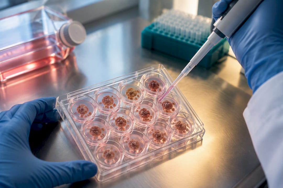
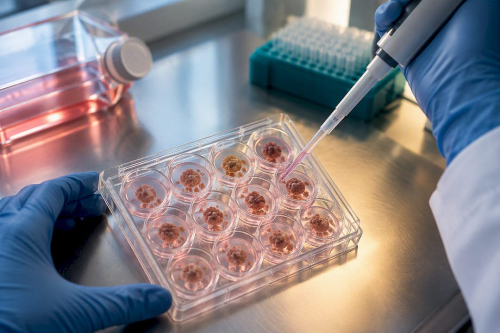
Organoids in cancer research are three-dimensional, stem-cell-derived tumor models that preserve a patient’s own histology and genetic fingerprint, and they are already showing real predictive value for some cancer therapies. The strongest evidence comes from large biobanks with documented histopathology and genomic concordance, and from a growing set of prospective trials. The honest caveat: reproducibility across labs, missing immune and vascular components, and multi-week turnaround times still limit how far these models can travel from the bench to the bedside today.
TL;DR:
- Organoids better replicate tumor architecture and heterogeneity than 2D cell lines, but they still lack immune and vascular components that influence drug response.
- Culturing tumor organoids requires precise documentation of reagent lot numbers, sample collection times, and culture conditions to ensure reproducibility across labs.
- Turnaround times from tissue collection to drug-response data typically span several weeks, limiting their use in rapidly progressing cancers without careful patient selection.
- Validating organoid fidelity relies on high histopathology and genomic concordance with the parent tumor, with drug response often matching patient outcomes above 80%.
- Standardization and shared biobank infrastructure are key to advancing organoid research into routine clinical practice, supported by increased funding and collaborative efforts.
Table of Contents
- What Are Organoids and How Do They Differ From Other Models?
- How Are Tumor Organoids Established and Validated?
- Where Do Organoids Actually Move the Needle in Cancer Research?
- What Evidence Supports Clinical Use of Patient-Derived Organoids?
- What Are the Real Limitations of Organoid Models?
- How Can Researchers Improve Organoid Reproducibility?
- Why HCRF Supports Organoid and Translational Cancer Research
- Our Perspective: What the Field Needs Next
- Sources
- FAQ
What Are Organoids and How Do They Differ From Other Models?
Organoids form when adult stem cells or induced pluripotent stem cells (iPSCs) are placed in an extracellular matrix scaffold, usually Matrigel or a similar basement membrane extract, and fed a cocktail of growth factors that mimics the signals those cells would receive inside a real organ. Left to their own devices, the cells self-organize. They divide, differentiate, and arrange themselves into three-dimensional structures that echo the architecture of the tissue they came from, crypts and villi in a colon organoid, ductal structures in a pancreatic one. That self-organizing property is the whole point: it is what separates an organoid from a simple ball of cells growing in a dish.
Patient-derived organoids (PDOs) start from a patient’s own tumor sample, so they carry that patient’s specific mutations, expression patterns, and drug sensitivities forward into the culture dish. iPSC-derived organoids start from reprogrammed skin or blood cells and are pushed down a developmental pathway toward a target tissue, useful for modeling hereditary cancer syndromes or studying tumor initiation from a normal starting point rather than an already-established tumor. Assembloids take this a step further by fusing two or more organoid types together, say, a tumor organoid with an immune-cell-containing organoid, to study how compartments interact. Spheroids, by contrast, are simpler: clusters of cells with no defined internal architecture, useful for basic viability screens but not for modeling structural complexity.
Why does any of this matter for cancer specifically? Because architecture and heterogeneity are not decoration. They are function.
- Two-dimensional cell lines flatten cells onto plastic, stripping away the 3D cell-to-cell contacts that shape drug response and gene expression.
- Patient-derived xenografts (PDX models) preserve architecture but require months to establish in mice, and they still filter the tumor through a non-human immune environment.
- Organoids sit in between: faster than PDX models, and closer to the source tumor’s actual biology than a flat monolayer of cells, which is one reason 3D organoid culture consistently outperforms 2D lines on physiological relevance, even as it costs more and runs at lower throughput.
How Are Tumor Organoids Established and Validated?
Everything downstream of an organoid experiment depends on what happens in the first hour after tissue leaves the operating room or biopsy suite. Surgical resections, needle biopsies, and malignant effusions (fluid drawn from the chest or abdomen) all serve as valid starting material, but they are not interchangeable. Resections yield more tissue and more cell types; biopsies are smaller but far more common in metastatic disease; effusions are often the only accessible sample in advanced cancers and can be surprisingly rich in viable tumor cells.
Cold ischemia time, the interval between when blood supply is cut off and when the tissue actually reaches culture media, is one of the most underappreciated variables in the entire workflow. Longer delays mean more cell death, more stress-response gene expression, and organoids that may no longer represent the tumor they came from.
Establishing a tumor organoid culture typically follows these steps:
- Mechanically and enzymatically dissociate the fresh tissue into small clusters or single cells.
- Embed the cell clusters in Matrigel or a comparable basement membrane extract, then plate them in submerged culture, air-liquid interface (ALI), or spinner-flask format depending on the tumor type and downstream assay.
- Feed the culture a defined growth-factor cocktail (commonly including EGF, R-spondin, Noggin, and Wnt3a depending on tissue of origin) and monitor for organoid formation over one to three weeks.
- Passage the organoids at defined intervals, tracking passage number carefully.
- Characterize the established line against the parent tumor using histology, targeted sequencing, and increasingly single-cell RNA sequencing, then run functional viability and drug-response assays to confirm the model behaves as expected.
Submerged Matrigel culture is the default choice for most epithelial tumors. Air-liquid interface formats work better when you need to preserve immune or stromal components alongside the tumor cells. Spinner or bioreactor culture suits scale-up when a lab needs large organoid numbers for high-throughput screening.
The reagents matter more than most protocols admit. Matrigel is a biologically derived product with lot-to-lot variability that can shift organoid growth rate and even drug-sensitivity readouts, and small changes to a growth-factor recipe or the timing of passaging can measurably alter gene expression. Any lab running organoid work needs a biosafety level appropriate to human tissue culture (typically BSL-2) and a documented chain of custody from operating room to incubator.
Pro Tip: Log your Matrigel lot number, passage number, and exact time-from-collection-to-culture for every organoid line. When a result looks off, these three variables are the first place to look, and reviewers increasingly expect them reported.
Where Do Organoids Actually Move the Needle in Cancer Research?
Drug screening is where organoids in cancer research have made the fastest and most visible impact. A single patient-derived organoid line can be split across a multi-well plate and exposed to dozens of compounds or combinations at a range of doses, producing dose-response curves that flag which agents actually shrink the organoid population versus which ones do nothing. Medium-throughput screens (tens to low hundreds of compounds) are now routine in academic cancer centers; true high-throughput screening (thousands of compounds) remains mostly the domain of specialized automated facilities, because organoids are pricier and slower to scale than flat cell lines.
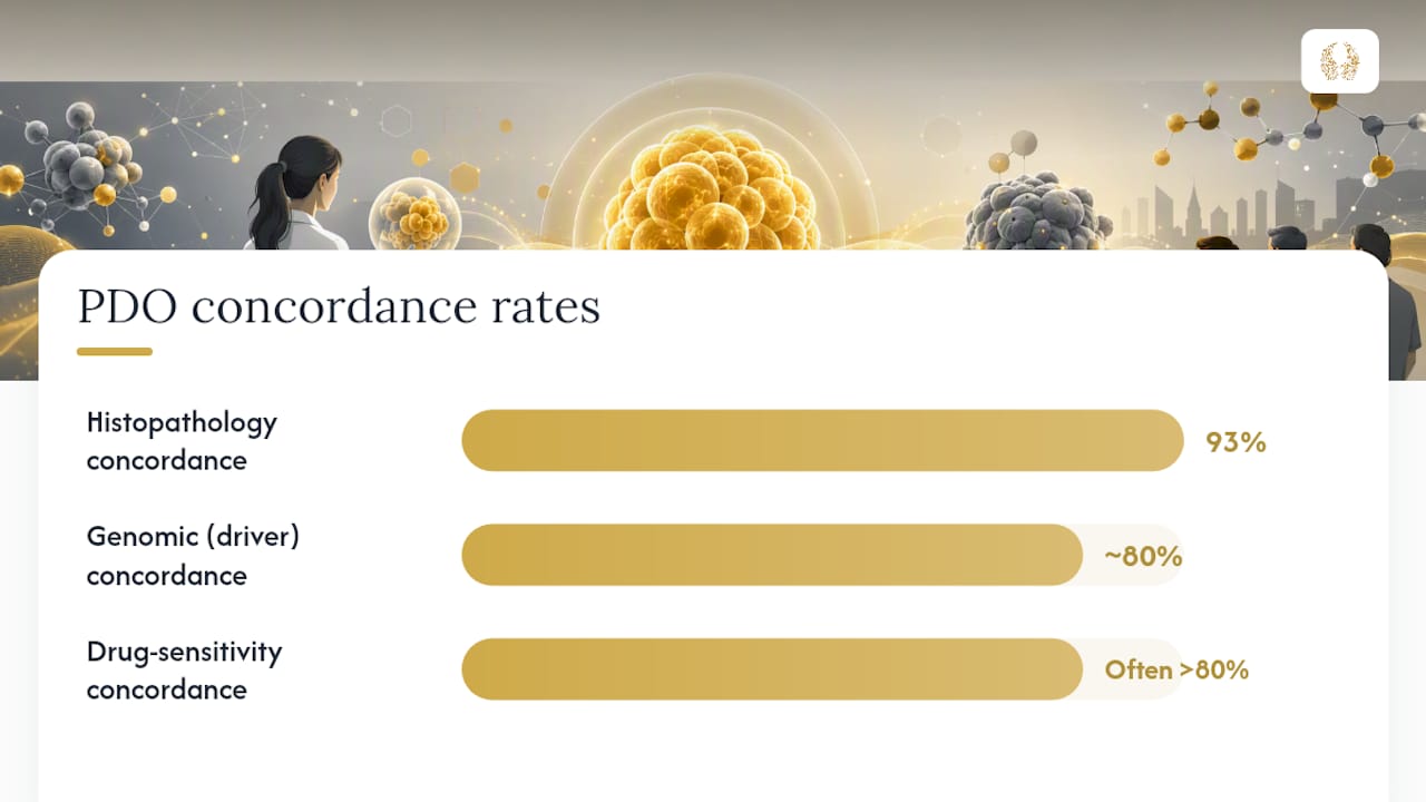
Across a pan-cancer organoid biobank spanning 220 organoid lines from 191 patients and 15 cancer types, histopathology concordance with the parent tumor reached 93%, with a median genomic concordance near 80% for driver mutations.
That kind of fidelity is what makes the “patient avatar” concept plausible: an organoid grown from a patient’s own tumor, tested against a therapy before that therapy is given to the patient. Reviews summarizing multiple prospective and retrospective studies report drug-sensitivity concordance often exceeding 80% between organoid response and actual patient outcome in selected cohorts, including metastatic colorectal, rectal, and gastric cancers, which is the closest thing the field currently has to a real-world validation signal for functional precision oncology.
Immuno-oncology applications add another layer. Co-culturing tumor organoids with a patient’s own T cells or with macrophages lets researchers watch, in real time, whether immune cells actually recognize and kill the tumor cells, and whether checkpoint inhibitors change that outcome. These systems have already linked macrophage-organoid signaling loops to drug resistance mechanisms, though they remain technically demanding and far from standardized across labs.
Beyond drug testing, organoid biobanks are proving useful for questions that have nothing to do with any single therapy:
- Tracking how subclones within a single tumor respond differently to the same drug, capturing intratumoral heterogeneity that a bulk tumor biopsy would average away.
- Modeling how cells acquire the mutations that let them seed a metastasis.
- Screening organoid panels for shared vulnerabilities, work that has already identified sensitivity to agents like talazoparib and combination partners capable of overcoming resistance in subsets of tumor models.
- Feeding organoid-derived genomic, transcriptomic, and functional drug-response data into multi-omics pipelines to nominate new biomarkers of treatment response.
What Evidence Supports Clinical Use of Patient-Derived Organoids?
The strongest data points come from large biobanking efforts that pair organoid generation with rigorous molecular comparison to the source tumor. The pan-cancer platform referenced above did not stop at concordance rates; it also reported a median gene expression correlation of 0.85 between organoids and their parent tumors, holding stable across passages, evidence that these models do not drift into some generic, unrepresentative state the longer they sit in culture.
Clinical translation is also visible in trial registrations. Review summaries put the number of PDO-related studies on ClinicalTrials.gov at roughly 30 trials in earlier surveys, spanning both observational biobanking efforts and interventional studies where organoid drug response actively guides treatment decisions.
| Evidence type | What it measures | Reported outcome |
|---|---|---|
| Pan-cancer PDO biobank | Histopathology concordance with parent tumor | 93% |
| Pan-cancer PDO biobank | Median genomic concordance (driver mutations) | ~80% |
| Pan-cancer PDO biobank | Median gene expression correlation | 0.85 |
| Prospective/retrospective clinical cohorts | Drug-sensitivity concordance with patient response | Often above 80% in selected cancers |
| ClinicalTrials.gov registrations | PDO-related trials (earlier review count) | ~30 trials |
Turnaround time is the practical wall standing between these encouraging numbers and everyday clinical decision-making. Generating and screening a PDO line often takes several weeks from sample collection to usable drug-response data, a window that works for many solid tumors but can outrun the clinical timeline in rapidly progressive cancers. Investigators need to document that turnaround honestly and triage which patients and sample types are realistic candidates for organoid-guided decisions rather than treating every case as eligible.
Data governance adds another layer clinicians cannot skip past. Because PDOs carry a patient’s living genetic material and can, in principle, be expanded and shared across institutions, consent processes need to spell out how long organoid lines will be stored, whether they will be shared with outside collaborators or biobanks, and whether future, not-yet-specified research uses are covered. Institutional review boards increasingly expect this level of detail before approving organoid-generation protocols tied to clinical care.
What Are the Real Limitations of Organoid Models?
Reproducibility problems trace back to the same reagent-sensitivity issues raised earlier, but at scale they become a field-wide credibility question rather than a single-lab inconvenience. Two labs running what looks like the identical published protocol can get organoids with meaningfully different drug-response profiles, because Matrigel lot, exact media formulation, and passage timing at the point of assay all shift outcomes in ways that standard method sections rarely capture in enough detail to reproduce.
The bigger structural gap is what standard PDOs leave out entirely.
- No stromal fibroblasts, which shape drug penetration and resistance in real tumors.
- No functional vasculature, so nutrient and drug delivery inside the organoid does not match delivery inside an actual tumor mass.
- No native immune infiltrate unless a co-culture system is deliberately added back in, which means a standard PDO drug screen can systematically miss therapies that work partly by recruiting or activating immune cells.
Cost and scale compound these biology gaps. Organoid culture demands more expensive reagents, more highly trained hands, and more time per data point than a 2D cell line assay, which is exactly why organoids still trail 2D systems on raw throughput even as they beat them on physiological relevance. Regulatory pathways for using organoid data in actual treatment decisions remain undefined in most jurisdictions, and cross-institutional data sharing runs into the same consent and governance questions raised in the clinical evidence section above.
Pro Tip: Treat every organoid drug-response result as hypothesis-generating until it has been checked against an orthogonal readout, ideally a genomic or histologic concordance check against the parent tumor, not as a stand-alone clinical answer.
How Can Researchers Improve Organoid Reproducibility?
Reproducibility is fixable, but only if labs agree to report the variables that actually move the needle rather than the variables that are easy to report.
- Document sample source, time from collection to culture, and cold ischemia duration for every organoid line, not just successful ones.
- Record Matrigel lot number, full media recipe, and passage number at the point each assay is run.
- Pair every drug-response experiment with a histologic and, where feasible, targeted genomic concordance check against the parent tumor.
- Run adequate biological replicates and a defined dose range for every compound, with pre-specified thresholds for what counts as a meaningful response before the data are collected, not after.
- Deposit characterization and drug-response data in shared biobank repositories using consistent metadata fields, so results from different institutions can actually be compared side by side.
- Reach for co-culture, organ-on-chip, or bioprinting approaches specifically when the research question depends on stromal, vascular, or immune interactions that a standard PDO cannot supply, rather than defaulting to the simplest model available.
Pro Tip: If your funding or publication timeline can absorb the added complexity, an organ-on-chip or immune co-culture add-on is usually worth it the moment your research question touches drug penetration, resistance mechanisms, or immunotherapy response, since a standard PDO cannot answer those questions on its own.
Why HCRF Supports Organoid and Translational Cancer Research
The Hippocratic Cancer Research Foundation is a 501©(3) nonprofit that funds the kind of “out of the box” cancer research that conventional grant cycles often pass over, work at the Robert H. Lurie Comprehensive Cancer Center of Northwestern University. Organoid platforms sit squarely in that category: promising, evidence-backed, and still too early-stage for many traditional funding pipelines to embrace at the pace the science deserves.
For researchers and students who want to go deeper on the biology behind these models, HCRF maintains educational resources covering the tumor microenvironment and the cell types that make up a tumor, both directly relevant to understanding what standard organoids capture and what they still miss.
- HCRF funds translational, high-risk cancer projects at a nationally recognized cancer center.
- Educational materials on tumor biology are freely available to researchers, clinicians, and students.
- Investigators interested in HCRF-funded translational work can explore ongoing innovative cancer research supported by the foundation.
Our Perspective: What the Field Needs Next
We think the next real leap in organoids in cancer research will not come from a flashier model. It will come from boring, unglamorous standardization: shared QC metrics, common reporting templates, and biobanks that talk to each other instead of sitting in institutional silos. HCRF believes funding should prioritize prospective validation studies and collaborative biobanking infrastructure over one-off proof-of-concept papers. That is where “out of the box” research earns its keep.
— HCRF
Support this kind of translational, evidence-driven cancer research directly. The foundation’s Cocktails for a Cure event kicks off a season of donor engagement that funds high-risk, high-reward projects often overlooked by traditional grants. For a larger-scale evening of giving, HCRF’s 13th Annual Wings To Cure Gala brings the community together to fund innovative research at the Robert H. Lurie Comprehensive Cancer Center. Every dollar raised moves projects like organoid biobanking from a hopeful hypothesis closer to a tool clinicians can actually trust.
This article is general information, not a substitute for advice from a qualified doctor. Consult a qualified healthcare professional about your own circumstances before acting on anything here.
Sources
- Patient-derived organoids across cancers reveal conserved tumor heterogeneity and actionable therapeutic vulnerabilities | Science Advances
- Cancer organoids: A platform in basic and translational research (PMC review)
- 3D organoids vs 2D cell lines in drug discovery and disease modeling | Biocompare
FAQ
Is Adenocarcinoma Hereditary?
Most adenocarcinomas arise from a combination of acquired mutations and environmental exposures rather than a single inherited gene, though hereditary syndromes such as Lynch syndrome and BRCA mutations raise risk for specific adenocarcinoma types. Organoid models derived from carriers of these syndromes are increasingly used to study how inherited mutations shape tumor behavior before cancer even develops.
What Cancer Was Cured in a Mouse?
Numerous cancer types, including certain leukemias, melanomas, and breast cancer models, have been successfully eliminated in mouse models using experimental therapies, but a cure in a mouse does not reliably predict a cure in humans. This gap is a major reason the field has shifted toward patient-derived organoids, which preserve human-specific tumor biology that mouse models cannot fully replicate.
What Are the Disadvantages of Organoids?
Standard organoid cultures lack the stromal, vascular, and immune components present in real tumors, which can bias drug-response results toward whatever a tumor cell alone would do. They are also costlier and slower to generate than 2D cell lines, prone to lab-to-lab variability from reagent lot differences, and typically take multiple weeks to produce usable results, a real constraint for rapidly progressing cancers.
What Organ Rarely Gets Cancer?
The heart is one of the organs where primary cancer is exceptionally rare, largely because heart muscle cells divide far less often than cells in tissues like the colon, lung, or breast, giving fewer opportunities for cancer-driving mutations to accumulate. Researchers have used organoid and stem-cell models to study why certain tissue types resist tumor formation, insight that could eventually inform prevention strategies elsewhere in the body.
Recommended

Discover the true cost of cancer research, from initial grants to full drug development. Learn how to make impactful donations today.
Read More →
Methodology guide for clinicians and trialists on basket trials in cancer: when to pool, expand, or confirm, plus pooled 18% ORR, design rules, and...
Read More →