
Tumor Cell Types: Cell Origins and Clinical Implications
July 27, 2026
Tumor Cell Types: Cell Origins and Clinical Implications

Tumors fall into three fundamental groups — benign, premalignant, and malignant — and the cell of origin within each group is what drives every clinical decision that follows. Malignant tumors are the ones properly called cancers: they invade surrounding tissue and can spread to distant sites, a capacity that benign tumors simply do not have. The major malignant cell classes are carcinomas, sarcomas, hematologic malignancies, CNS tumors, germ cell tumors, neuroendocrine tumors, and melanocytic tumors — each arising from a distinct cell lineage and each demanding a different diagnostic and treatment approach.
- Benign and premalignant lesions are managed locally (surveillance, excision) because they lack the capacity for systemic spread.
- Malignant tumors require staging, and often systemic therapy, because invasion and metastasis define them.
- Cell of origin — confirmed by histology, immunohistochemistry, and molecular profiling — is the single most important factor in choosing targeted therapy and predicting prognosis.
Table of Contents
- What is a tumor, and how does it differ from cancer?
- Benign, premalignant, and malignant: how the three categories behave
- Major tumor cell types by tissue and cell of origin
- What makes malignant cells different at the cellular level?
- How tumor cell type is established through diagnosis
- Why tumor cell type shapes prognosis and treatment
- The tumor microenvironment and intratumor heterogeneity
- Key Takeaways
- Why classification accuracy is the foundation of everything we fund
- How HCRF supports the research that makes better classification possible
- Useful sources and further reading
What is a tumor, and how does it differ from cancer?
A tumor, or neoplasm, is a mass of abnormal cells that grow beyond normal tissue boundaries and serve no physiological function. That definition covers a wide range: a benign lipoma on the back and a metastatic pancreatic adenocarcinoma are both technically tumors. Cancer is the narrower term — it applies only when the tumor is malignant, meaning its cells can invade adjacent structures and seed distant organs.
That distinction carries immediate clinical weight. A benign tumor found on imaging may need nothing more than watchful waiting; a malignant one triggers a staging workup, a multidisciplinary tumor board review, and often systemic therapy. Cleveland Clinic’s patient resource notes that many tumors are benign and clarifies when lesions require treatment versus surveillance — a reminder that not every mass is an emergency, but none should be dismissed without proper evaluation.
Why the distinction matters clinically:
- A benign tumor that compresses a nerve or gland may still need surgical removal even when malignancy is ruled out.
- Premalignant lesions (dysplasia, carcinoma in situ) require close surveillance because they carry measurable transformation risk.
- Malignant tumors demand staging to determine whether systemic therapy is needed alongside local treatment.
- Prognosis and five-year survival estimates are tied directly to whether a tumor is malignant and, if so, to its cell type and grade.
Pro Tip: When a radiologist describes a lesion as “indeterminate,” that is the clinical signal for biopsy — not reassurance. Watchful waiting is appropriate only after tissue has confirmed a benign diagnosis.
Benign, premalignant, and malignant: how the three categories behave
| Category | Typical Behavior | Representative Examples |
|---|---|---|
| Benign | Slow growth, well-circumscribed, no invasion or metastasis | Lipoma, adenoma, leiomyoma, angiofibroma |
| Premalignant | Abnormal cells with transformation potential; no invasion yet | Dysplasia, carcinoma in situ, Barrett’s esophagus |
| Malignant | Invasive growth, metastatic potential, disrupts normal architecture | Adenocarcinoma, squamous cell carcinoma, osteosarcoma |
MedlinePlus notes there are over 100 distinct cancer types, each named for its tissue of origin and cell type — which is why the category table above is only the beginning of the story. Borderline and ambiguous lesions add real complexity to this tidy three-tier model.
Clinically ambiguous situations to know:
- Carcinoma in situ (CIS): Malignant cells confined to the epithelium with no basement membrane breach. Treated as malignant in most protocols.
- Low-grade dysplasia: Monitored closely; transformation rates vary by organ site.
- Borderline ovarian tumors: Neither clearly benign nor frankly malignant; managed with fertility-sparing surgery in appropriate patients.
- Gastrointestinal stromal tumors (GIST): Risk stratified by mitotic rate and size rather than a simple benign/malignant label.
Major tumor cell types by tissue and cell of origin
Carcinomas account for the majority of human cancers, making them by far the most common malignant cell class. Sarcomas, hematologic malignancies, and the remaining classes are rarer but no less clinically significant. The table below maps each major class to its defining features.
| Cell Class | Cell of Origin | Key Markers | Common Primary Sites | Typical Spread |
|---|---|---|---|---|
| Carcinoma | Epithelial cells | Cytokeratins, EpCAM, CDH1 | Breast, lung, colon, prostate, pancreas | Lymph nodes, liver, lung, bone |
| Sarcoma | Mesenchymal cells | Vimentin, SMA, S100 (MPNST) | Bone, soft tissue, retroperitoneum | Lung (hematogenous) |
| Leukemia/Lymphoma | Hematopoietic cells | CD3, CD20 | Bone marrow, lymph nodes, spleen | Systemic (blood/marrow) |
| Multiple Myeloma | Plasma cells | CD138, MZB1, JCHAIN, SDC1 | Bone marrow | Bone, kidney |
| CNS Tumors | Glial/neuronal cells | GFAP, IDH1/2, OLIG2 | Brain, spinal cord | Local infiltration (rarely systemic) |
| Germ Cell Tumors | Primordial germ cells | AFP, hCG, OCT4, PLAP | Gonads, mediastinum, retroperitoneum | Lymph nodes, lung, liver |
| Neuroendocrine Tumors | Neuroendocrine cells | Chromogranin A, Synaptophysin, Ki-67 | Pancreas, GI tract, lung | Liver, bone |
| Melanoma | Neural crest melanocytes | S100, MITF, HMB-45 | Skin, uvea, mucosa | Lung, brain, liver, skin |
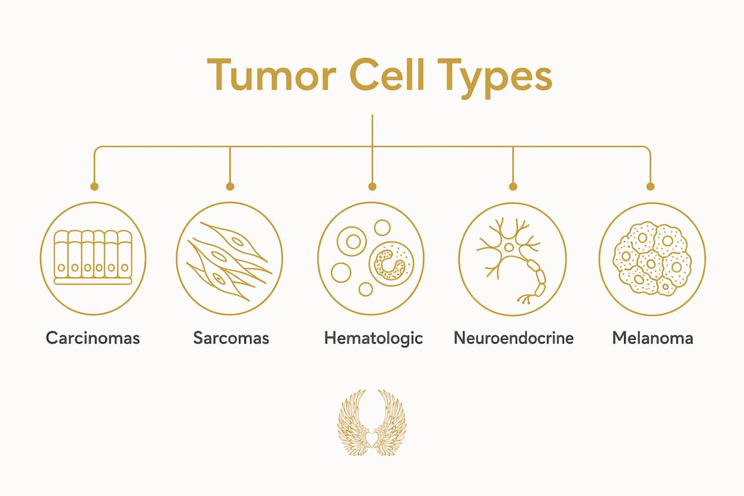
Carcinomas
Carcinomas split into two major subtypes: adenocarcinomas, which arise in glandular epithelium (breast, colon, lung, prostate), and squamous cell carcinomas, which originate in stratified squamous epithelium (head and neck, cervix, skin, esophagus). Small-cell carcinoma of the lung is a particularly aggressive variant — neuroendocrine in phenotype, it grows rapidly and responds initially to platinum-based chemotherapy but almost always recurs.

Sarcomas
Sarcomas arise from mesenchymal tissue and tend to spread hematogenously to the lung rather than through lymphatics. Osteosarcoma peaks in adolescents; leiomyosarcoma and liposarcoma are more common in adults. High-grade sarcomas carry a poor prognosis without wide surgical resection combined with chemotherapy.

Hematologic malignancies
Leukemias are “liquid” cancers of the bone marrow; lymphomas are solid tumors of lymphoid tissue. The presence of Reed-Sternberg cells on biopsy distinguishes Hodgkin lymphoma from Non-Hodgkin lymphoma — a distinction that changes treatment entirely. Multiple myeloma, arising from malignant plasma cells, is confirmed by CD138 positivity and the detection of a monoclonal protein.
CNS tumors, germ cell tumors, neuroendocrine tumors, and melanoma
Gliomas infiltrate brain parenchyma and are graded by IDH mutation status and 1p/19q codeletion under the current WHO classification. Germ cell tumors are among the most chemotherapy-sensitive solid tumors — testicular seminoma has a cure rate exceeding 95% with platinum-based regimens. Neuroendocrine tumors range from indolent well-differentiated carcinoids to highly aggressive large-cell neuroendocrine carcinomas, stratified by Ki-67 proliferation index. Melanoma, arising from neural crest-derived melanocytes, carries the highest metastatic potential of any skin malignancy; see our guide on moles versus melanoma for practical clinical differentiation.
Notable aggressive subtypes to recognize:
- Small-cell lung carcinoma: rapid doubling time, early metastasis, paraneoplastic syndromes
- High-grade osteosarcoma: peak incidence in adolescents, requires neoadjuvant chemotherapy
- Glioblastoma (WHO grade 4): median survival under 15 months despite multimodal therapy
- Burkitt lymphoma: one of the fastest-growing human tumors, driven by MYC translocation
What makes malignant cells different at the cellular level?
The cellular hallmarks of malignancy — first systematized by Hanahan and Weinberg — describe the biological capabilities cancer cells must acquire to become fully malignant. Understanding them helps explain why some tumors are aggressive and why certain therapies work.
Core hallmarks of malignant tumor cells:
- Sustained proliferative signaling: Oncogene activation (KRAS, EGFR, HER2) drives growth independent of external signals.
- Resisting cell death: Mutations in TP53 and overexpression of BCL-2 family proteins disable apoptosis.
- Replicative immortality: Telomerase reactivation prevents chromosomal shortening and allows unlimited division.
- Inducing angiogenesis: VEGF secretion recruits new blood vessels to feed the growing tumor mass.
- Activating invasion and metastasis: Matrix metalloproteinases degrade the extracellular matrix; epithelial-mesenchymal transition (EMT) strips cells of adhesion molecules and confers migratory capacity.
- Evading immune destruction: PD-L1 overexpression and MHC class I downregulation shield tumor cells from cytotoxic T cells.
- Deregulating cellular energetics: The Warburg effect (aerobic glycolysis) fuels rapid proliferation even in low-oxygen environments.
- Genomic instability: Defects in DNA repair (mismatch repair, BRCA1/2) accelerate mutation accumulation and drive tumor evolution.
EMT deserves special attention. When carcinoma cells lose epithelial markers like EpCAM and CDH1 while gaining mesenchymal markers like vimentin and fibronectin, they become invasive and enter the bloodstream as circulating tumor cells. That transition is also what makes them harder to detect by standard epithelial marker panels in single-cell analyses.
Pro Tip: Pathologists infer aggressive behavior from a combination of high mitotic index, geographic necrosis, and loss of TP53 expression — no single feature is sufficient. A tumor with all three warrants urgent multidisciplinary review.
How tumor cell type is established through diagnosis
Establishing cell type follows a stepwise pathway. Imaging identifies the lesion; tissue sampling provides the material; histology, immunohistochemistry (IHC), and molecular profiling together assign the diagnosis.
Common IHC markers and what they reveal:
- ER/PR/HER2 (breast carcinoma): determines eligibility for hormone therapy and trastuzumab
- EGFR/ALK/ROS1 (lung adenocarcinoma): identifies patients for tyrosine kinase inhibitors
- CD20 (B-cell lymphoma): confirms rituximab eligibility
- IDH1 R132H (glioma): prognostic and now a WHO classification criterion
- BRAF V600E (melanoma, colorectal): targets vemurafenib and encorafenib-based regimens
- Chromogranin A / Synaptophysin (neuroendocrine tumors): confirms neuroendocrine lineage
Diagnostic workflow:
- Imaging (CT, MRI, PET-CT): characterize lesion size, location, and suspicious features.
- Tissue sampling (core needle biopsy, excisional biopsy, or endoscopic sampling): obtain adequate material for analysis.
- Histopathology (H&E staining): assess architecture, nuclear grade, mitotic index, and necrosis.
- Immunohistochemistry: confirm cell lineage and identify therapeutic targets.
- Molecular profiling (NGS, FISH): detect driver mutations, gene fusions, and copy-number alterations when targeted therapy is being considered.
- Single-cell sequencing or pan-cancer classifiers (research and select clinical settings): resolve cell identity in heterogeneous or metastatic samples of unknown primary.
The scATOMIC pan-cancer classifier, trained on over 300,000 single cells, demonstrated high accuracy in identifying cancer and non-malignant cell types across multiple tumor types — including predicting primary origin in metastatic samples where histology alone was ambiguous.
Why tumor cell type shapes prognosis and treatment
Cell type is not just a label — it is the roadmap for every treatment decision. Understanding how staging relates to cell type is equally important: grade reflects how differentiated the cells are, while stage reflects how far the tumor has spread.
Treatment pathways by tumor class:
- Localized benign tumors: Observation or surgical excision; no systemic therapy needed.
- Carcinomas: Surgery combined with chemotherapy, radiation, and increasingly targeted agents or immunotherapy based on molecular subtype.
- Hematologic malignancies: Primarily systemic therapy (chemotherapy, immunotherapy, CAR-T); transplant in selected cases.
- Sarcomas: Wide surgical resection is the cornerstone; chemotherapy added for high-grade or metastatic disease.
- Germ cell tumors: Platinum-based chemotherapy with curative intent even in metastatic disease.
Two clinical examples show how cell type changes everything. Small-cell lung carcinoma (SCLC) is treated with platinum-etoposide chemotherapy and immunotherapy from the outset — surgery plays almost no role because SCLC is nearly always systemic at diagnosis. Non-small-cell lung carcinoma (NSCLC), by contrast, is first evaluated for EGFR, ALK, and ROS1 alterations; patients with driver mutations receive oral targeted agents and can achieve durable responses measured in years rather than months.
Molecular subtypes within carcinomas add another layer. In breast cancer, hormone receptor-positive (ER+/PR+) tumors respond to endocrine therapy; HER2-amplified tumors respond to anti-HER2 agents; triple-negative breast cancer (TNBC) lacks all three targets and relies on chemotherapy and immunotherapy. That single biopsy result redirects the entire treatment plan.
“The malignant tumor’s ability to invade and metastasize — not just rapid growth — is what dictates the need for systemic therapy and aggressive management.” — NCBI Bookshelf, The Development and Causes of Cancer
Cancer stem cells add yet another dimension. These tumor-initiating cells often resist standard chemotherapy and are thought to drive relapse; identifying stem-like transcriptional programs is an active research priority with direct implications for targeted clinical trials.
The tumor microenvironment and intratumor heterogeneity
A tumor is not a uniform mass of identical malignant cells. It is an ecosystem — cancer cells surrounded by immune cells, fibroblasts, endothelial cells, and extracellular matrix, all communicating through cytokines and growth factors. That multicellular community, the tumor microenvironment (TME), increasingly predicts outcomes more accurately than histology alone.
The EcoTyper framework, applied across multiple types of human carcinoma and thousands of tumors, identified several clinically distinct multicellular communities — including three with myeloid and stromal elements linked to adverse survival. That finding means two tumors with identical histology can have radically different immune contexts and, therefore, different responses to immunotherapy.
Intratumor heterogeneity compounds the challenge. Different regions of the same tumor can harbor distinct genetic subclones, meaning a single biopsy may not capture the full picture. Single biopsies can under-sample heterogeneous tumors; multidisciplinary tumor boards and repeat sampling or molecular assays reduce that sampling error in practice.
“The tumor microenvironment is increasingly used to stratify patients because multicellular communities and cell states often predict outcomes more accurately than histology alone.” — Nature Communications, scATOMIC pan-cancer classifier study
When genomic instability is low — in diploid tumors — copy-number variation methods may fail to detect malignant cells at all. Marker-free transcriptional classifiers or combined proteogenomic approaches become necessary in those cases, a limitation that standard clinical pathology workflows do not yet routinely address.
Emerging tools reshaping classification:
- scATOMIC: Pan-cancer single-cell classifier trained on >300,000 cells; resolves immune subtypes and rare populations.
- EcoTyper: Identifies transcriptionally defined cell states and multicellular communities from bulk and single-cell data.
- InferCNV / scMalignantFinder: Detect copy-number alterations in single-cell data to separate malignant from normal cells.
- Spatial transcriptomics: Maps cell states to physical tumor locations, revealing how communities are organized in tissue.
Key Takeaways
Tumor cell type — determined by cell of origin, histology, and molecular profiling — is the single most consequential variable in cancer diagnosis, prognosis, and treatment selection.
| Point | Details |
|---|---|
| Tumor vs. cancer | Only malignant tumors capable of invasion and metastasis are properly called cancers; benign tumors remain localized. |
| Cell-of-origin classes | Carcinomas, sarcomas, hematologic malignancies, CNS tumors, germ cell, neuroendocrine, and melanoma each require distinct diagnostic and treatment approaches. |
| Molecular subtypes matter | Markers like ER/PR/HER2 in breast cancer and EGFR/ALK in lung adenocarcinoma redirect the entire treatment plan based on a single biopsy result. |
| Microenvironment and heterogeneity | The tumor ecosystem and intratumor heterogeneity often predict outcomes more accurately than histology alone; single biopsies can miss critical subclones. |
| HCRF’s role | The Hippocratic Cancer Research Foundation funds the kind of molecular diagnostics and single-cell research that makes precise classification — and better treatment — possible. |
Why classification accuracy is the foundation of everything we fund
From where we stand at the Hippocratic Cancer Research Foundation, the science of tumor cell classification is not an abstract academic exercise. It is the foundation on which every treatment advance is built. When a researcher can precisely identify which cell gave rise to a tumor — and map the molecular alterations that drove its transformation — the path to a targeted therapy becomes visible. Without that precision, we are treating shadows.
We believe the most urgent investments in cancer research right now are in the tools that sharpen that precision: single-cell transcriptomics, pan-cancer molecular classifiers, and the translational work that moves those tools from research labs into clinical pathology workflows. Donor-funded research at partner cancer centers, including the Robert H. Lurie Comprehensive Cancer Center of Northwestern University, has supported exactly these kinds of projects — work that is quietly rewriting what we know about how tumors are organized and how they should be treated.
The gap between what we know in the lab and what reaches patients is still too wide. Closing it requires sustained, courageous funding of research that does not always follow the conventional path. That is precisely the kind of work HCRF was built to support.
How HCRF supports the research that makes better classification possible
The science in this article — single-cell classifiers, molecular profiling, microenvironment mapping — does not happen without dedicated funding. The Hippocratic Cancer Research Foundation exists to make sure the most promising, unconventional cancer research gets the support it deserves.

HCRF funds diagnostics research, single-cell studies, and clinical translation projects at the Robert H. Lurie Comprehensive Cancer Center of Northwestern University. Every dollar we raise goes toward closing the gap between laboratory discovery and patient care. If the science of tumor cell classification matters to you — if you believe that precision in diagnosis is the first step toward a cure — we invite you to be part of this mission.
Learn more about our work and support cancer research that is changing what is possible. Visit the Hippocratic Cancer Research Foundation to explore our current projects and find out how your support drives real impact.
Useful sources and further reading
- The Development and Causes of Cancer — NCBI Bookshelf: Foundational chapter covering the benign-vs-malignant distinction, invasion, metastasis, and the major cell-of-origin classes; the primary authority for the tumor-vs-cancer definition used throughout this article.
- Cancer Classification — SEER Training (NCI): The international standard for histological cancer classification, including the ICD-O-3 framework; covers carcinoma, sarcoma, myeloma, leukemia, lymphoma, and mixed types with prevalence data.
- Pan-Cancer Classification of Single Cells in the Tumour Microenvironment — Nature Communications: Describes the scATOMIC classifier trained on >300,000 cells; landmark paper for understanding single-cell approaches to tumor cell identification and microenvironment analysis.
- Identification of Malignant Cells in Single-Cell Transcriptomics Data — PMC: Comprehensive review of computational approaches to distinguishing malignant from non-malignant cells in scRNA-seq data; covers EMT, copy-number inference, and marker-based strategies.
- Atlas of Clinically Distinct Cell States and Ecosystems Across Human Solid Tumors — PMC: Introduces EcoTyper and the concept of carcinoma ecotypes; identifies 10 multicellular communities with prognostic significance across 16 cancer types.
- Tumor (Neoplasm): Types, Symptoms & Treatment — Cleveland Clinic: Patient-facing clinical resource explaining when tumors require treatment versus surveillance; practical framing for the benign-vs-malignant distinction.
- Cancer Overview — MedlinePlus: Accessible public health overview noting over 100 cancer types and the naming convention by tissue of origin; useful entry point for non-specialist readers.
- Single-Cell Tumor Cluster Classification Review: Practitioner-level review of threshold-based cluster classification in single-cell analyses; addresses heterogeneity, sampling error, and the >60% tumor-cell threshold used in tools like SCANER.
- A to Z List of Cancer Types — National Cancer Institute: Comprehensive NCI index of cancer types by histology and primary site; authoritative reference for the full scope of tumor diversity.
- Hippocratic Cancer Research Foundation: HCRF’s organizational home page describing its mission, funded research programs at Northwestern’s Lurie Cancer Center, and ways to support translational cancer research.
Recommended
- Hippocratic Cancer Research Foundation: Innovative Therapies | 4 Types of Malignant Tumors: What You Need to Know
- Hippocratic Cancer Research Foundation: Innovative Therapies | Why Are Some Cancers More Common Than Others?
- Hippocratic Cancer Research Foundation: Innovative Therapies | T2 N0 Cancer Staging: What It Means for You
- Hippocratic Cancer Research Foundation: Innovative Therapies | What Causes Cancer: Key Factors and Prevention

Practical steps to build peer-to-peer cancer campaigns, protect donors from deceptive charities, and use HCRF resources for clear stewardship and...
Read More →