


Early Detection Biomarkers: What Clinicians Need to Know
August 21, 2026
Early Detection Biomarkers: What Clinicians Need to Know

Early detection biomarkers are measurable molecules, such as circulating tumor DNA (ctDNA), methylation patterns, proteins, microRNAs, and exosomes, shed into blood or other fluids that flag malignancy or precursor lesions before symptoms appear. The honest clinical answer for 2026 is this: several single-cancer tests have earned regulatory clearance and belong in standard practice, but multi-cancer early detection (MCED) panels remain largely investigational and are not substitutes for guideline-based screening.
The evidence base is moving fast, but it hasn’t closed the gap between promising signal and proven benefit. A systematic review of blood-based multi-cancer screening tests covering 20 studies and more than 109,000 people found no completed controlled trial demonstrating that MCEDs reduce cancer mortality. The NHS-Galleri trial and the Early Detection Research Network (EDRN) are working to close exactly that gap, one of the largest prospective efforts testing whether a positive signal actually changes outcomes rather than just flagging disease earlier on paper.
Here’s what matters for practice right now:
- Single-analyte, FDA-cleared tests like Cologuard have a defined screening role and years of validation behind them.
- MCED and pan-cancer liquid biopsy panels are mostly Laboratory Developed Tests (LDTs), meaning they haven’t cleared the same regulatory bar as approved screening devices.
- Stage I sensitivity is still the field’s weak point. Even strong composite biomarker panels report sensitivities around 55 to 57 percent for stage I disease, which means a negative result never rules out early cancer.
Pro Tip: Treat a positive MCED signal as a probability update, not a diagnosis. Route it through risk-scoring, then organ-directed imaging, then targeted tissue diagnostics, rather than jumping straight to invasive workup.
Key Takeaways
Early detection biomarkers show strong analytical promise, but only FDA-approved single-analyte tests like Cologuard currently meet the evidentiary bar for population screening, while MCED panels remain investigational adjuncts pending mortality-outcome trials.
| Point | Details |
|---|---|
| Know the regulatory tier | Cologuard is FDA-approved for screening; most MCED panels operate as LDTs with variable validation rigor. |
| Stage I sensitivity is the bottleneck | Even strong panels report sensitivity around 55 to 57 percent for stage I disease. |
| Specificity threshold matters most | Screening-grade claims require sensitivity reported at a fixed specificity of 98 percent or higher. |
| Treat positive results as probability, not diagnosis | Route findings through imaging and multidisciplinary review before invasive workup. |
| Demand prospective, external validation | Case-control accuracy claims routinely overestimate real-world performance. |
Table of Contents
- What Are the Major Classes of Early Detection Biomarkers?
- What Distinguishes Screening Biomarkers From Diagnostic Ones?
- How Do Multi-Cancer Early Detection Tests Work?
- What Analytical Methods Detect Low-Frequency Tumor Signals?
- How Should Clinical Studies on Biomarkers Be Evaluated?
- What Are the Limitations and Risks of Biomarker Testing?
- How Should a Positive Biomarker Result Be Interpreted Clinically?
- How Should Researchers Design and Validate New Biomarkers?
- What Recent Research Directions Show the Most Promise?
- What Is the Current Regulatory Status of These Tests?
- Why This Research Matters to HCRF
- Frequently Asked Questions
- Sources
What Are the Major Classes of Early Detection Biomarkers?
Each biomarker class exploits a different biological leak from a tumor, and each comes with its own sensitivity ceiling at low tumor burden. Understanding the mechanism behind each one tells you why a test performs well in one cancer type and poorly in another.
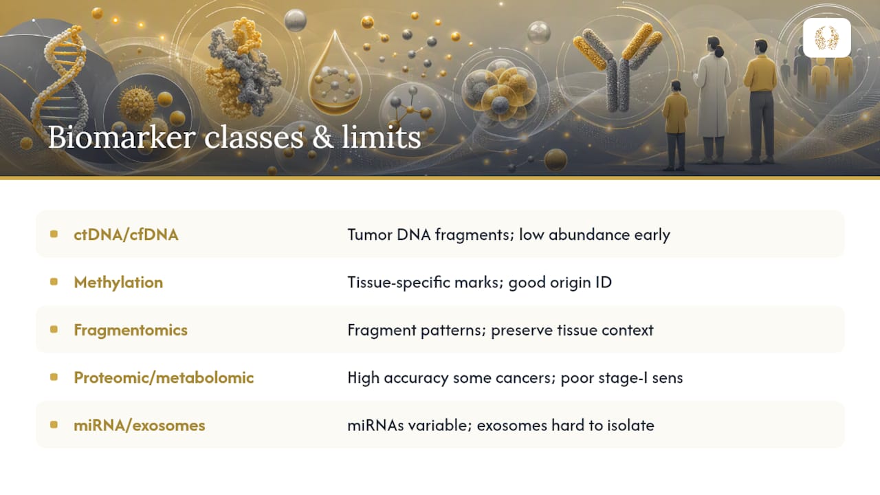
Circulating tumor DNA (ctDNA) and cell-free DNA (cfDNA) are fragments of DNA shed by dying tumor cells into the bloodstream. Assays detect tumor-specific mutations, copy number changes, or structural variants against a background of normal cfDNA. The core limitation is abundance: early-stage tumors shed vanishingly little ctDNA relative to their volume, so a mutation-only readout at ultra-low variant allele fractions requires deep sequencing and aggressive error correction to avoid drowning in background noise.
Methylation patterns capture epigenetic marks, chemical tags on DNA that regulate gene expression, and these marks are often tissue-specific. That property makes methylation profiling unusually good at tissue-of-origin prediction, which is why it anchors most current MCED platforms. Cologuard, the FDA-cleared colorectal screening test, relies partly on stool DNA methylation markers, an approach with over a decade of accumulated validation data.
Fragmentomics analyzes the physical characteristics of cell-free DNA fragments, their length distribution, end motifs, and nucleosome positioning, rather than their sequence. Because tumor-derived fragments carry a distinct chromatin signature, fragmentomic features preserve tissue context even when mutation signal is too sparse to call reliably. That makes fragmentomics attractive for tumor-naive population screening, where you don’t know in advance which mutation to look for.
Proteomic and metabolomic markers measure circulating proteins and small-molecule metabolites altered by tumor metabolism or the surrounding immune response. These panels can achieve striking accuracy for some cancers; a 2026 study reported composite proteomic and metabolomic biomarkers with an AUC of 0.97 for ovarian cancer and 0.91 for lung cancer, though stage I sensitivity lagged well behind those headline numbers.
MicroRNAs (miRNAs) and exosomes round out the panel. MiRNAs are short regulatory RNA molecules that circulate stably in blood, but inter-patient variability complicates setting a universal cutoff. Exosomes, tiny vesicles that tumor cells release carrying protein and nucleic acid cargo, are biologically rich but technically hard to isolate at scale, a bottleneck a recent review on emerging early-detection biomarkers flags as a major barrier to clinical standardization.
Circulating tumor cells (CTCs) are whole cancer cells shed into the bloodstream. They’re rare, often fewer than 10 cells per 10 milliliters of blood in early disease, which limits their use for detection even though they’re valuable for monitoring established disease.
A few practical notes on specimen handling: blood remains the dominant matrix for MCED work, but urine and saliva are gaining traction for organ-specific tests where local shedding concentrates the signal. Pre-analytic handling matters enormously; delayed plasma separation lets white blood cells lyse and release genomic DNA that dilutes true ctDNA signal, an error that can silently degrade sensitivity before the sample ever reaches the sequencer.
Statistic Callout: Multi-class biomarker panels that combine methylation, protein, and aneuploidy signals report overall sensitivities of roughly 53 to 61 percent at specificities above 98 percent, a meaningful gain over single-analyte tests in the same validation sets.
Pro Tip: No single biomarker class captures everything a tumor sheds. Combining a tumor-fraction signal (ctDNA mutations) with a tissue-context signal (methylation or fragmentomics) improves both sensitivity and tissue-of-origin accuracy more than doubling down on either signal alone.
What Distinguishes Screening Biomarkers From Diagnostic Ones?
A biomarker validated for one purpose is not automatically valid for another, and conflating these categories is one of the most common misreadings of the literature. Four distinct roles exist, and each demands its own evidence standard.
Screening biomarkers are meant for asymptomatic, average-risk populations. Because disease prevalence is low in that setting, even a test with strong sensitivity can generate an overwhelming number of false positives unless specificity is extremely high, typically above 98 to 99 percent. Screening claims also require proof of clinical benefit: a reduction in cancer mortality or in advanced-stage diagnoses, not just earlier detection on paper.
Diagnostic biomarkers confirm or rule out disease in a patient who already has symptoms or an abnormal finding. The prevalence in that population is much higher, so the same specificity threshold that would flood a screening program with false positives is often perfectly adequate diagnostically.
Prognostic biomarkers predict how disease will behave regardless of treatment, useful for staging and counseling. Predictive biomarkers forecast whether a specific treatment will work. The distinction matters clinically: establishing a predictive claim requires knowing biomarker status across both treated and untreated groups and testing for a treatment-by-biomarker interaction, not simply observing that biomarker-positive patients do better. A biomarker that’s prognostic in observational cohorts gets misapplied constantly as if it were predictive, or worse, as if it were screening-ready.
| Evaluation dimension | What to look for in the paper |
|---|---|
| Intended use | Screening, diagnostic, or minimal residual disease (MRD) monitoring, stated explicitly |
| Sensitivity by stage | Reported separately for stage I/II, not pooled across all stages |
| Specificity | Fixed threshold, ideally at or above 98 percent for screening claims |
| Clinical evidence level | Case-control, prospective cohort, or randomized trial |
Pro Tip: When a paper reports “sensitivity at 98 percent specificity” rather than an unqualified sensitivity figure, that’s a sign the authors understand screening-grade reporting. A bare sensitivity number without a matched specificity threshold is close to meaningless for population screening.
How Do Multi-Cancer Early Detection Tests Work?
MCED tests fall into a few architectural camps, and the differences shape what each one is realistically good for. Mutation-only panels sequence targeted gene regions for tumor-specific variants. Methylation-based panels, the dominant architecture in current commercial and research MCED platforms, profile thousands of methylation sites to generate both a cancer signal and a tissue-of-origin prediction. Multimodal platforms combine DNA signals with circulating proteins, aiming to catch cancers that shed little ctDNA but plenty of protein.
A few names anchor the literature here. CancerSEEK was an early proof-of-concept multimodal test combining mutation detection with protein biomarkers across eight cancer types, useful mainly as a demonstration that a single blood draw could plausibly flag multiple cancers, not as a clinical product. Galleri, from GRAIL, is a methylation-based MCED test currently marketed as an LDT and under evaluation in the NHS-Galleri trial, one of the largest prospective efforts to see whether population screening with this approach actually shifts stage at diagnosis. Guardant Health offers ctDNA-based assays spanning screening research and minimal residual disease monitoring, illustrating how the same underlying technology serves different clinical questions depending on the assay’s design. Cologuard, from Exact Sciences, remains the clearest precedent for what regulatory success looks like: an FDA-approved, stool-based methylation and DNA test for colorectal screening with over a decade of accumulated outcomes data. The Early Detection Research Network, funded by the National Cancer Institute, functions as the field’s validation infrastructure, running the multisite studies and biospecimen banking that individual test developers can’t easily replicate alone.
The regulatory distinction here isn’t a technicality. FDA-approved tests like Cologuard went through premarket review with a defined clinical validation bar; most MCED tests, by contrast, run as LDTs, which follow a different regulatory pathway and don’t carry the same premarket evidence requirement. That doesn’t mean LDTs are unreliable, but it does mean the validation rigor varies test to test, and clinicians ordering one should ask for the underlying data rather than assuming FDA-equivalent scrutiny.
Statistic Callout: Large MCED feasibility studies referenced in the CCGA and related landscape reviews consistently show specificity above 98 percent is achievable, but sensitivity still tracks tightly with cancer stage. Detection rates climb sharply from stage I to stage IV across nearly every cancer type these panels cover.
Pro Tip: Read the cancer-type breakdown, not the headline sensitivity number. MCED panels tend to perform well on cancers that already shed abundant ctDNA (pancreatic, liver, ovarian) and poorly on those that don’t (prostate, thyroid). A single blended sensitivity figure hides that variation.
What Analytical Methods Detect Low-Frequency Tumor Signals?
Finding a tumor mutation present at one part in ten thousand cell-free DNA molecules is a signal-processing problem as much as a biology problem, and the technology stack reflects that.
Error suppression is the foundation. Standard next-generation sequencing (NGS) introduces enough error to swamp true low-frequency variants, so modern ctDNA assays use unique molecular identifiers (UMIs), short barcode sequences tagged onto each DNA molecule before amplification, to distinguish real mutations from PCR and sequencing artifacts. Duplex sequencing takes this further by independently tagging both DNA strands, pushing error rates low enough to call variants at allele fractions below 0.1 percent.

Digital PCR (dPCR) partitions a sample into thousands of tiny reactions, each amplifying independently, which gives extremely sensitive and precise quantification for a small number of known mutations. It’s fast and cheap but doesn’t scale to the genome-wide discovery work MCED requires. Shallow whole-genome sequencing trades depth for breadth, useful for detecting copy number changes across the genome. Targeted methylome sequencing sits in between, sequencing thousands of specific methylation sites deeply enough to build a robust tissue-of-origin classifier, which is why it dominates current MCED architecture.
Emerging platforms are starting to reach clinical pipelines. Nanobiosensors, devices that detect biomolecules through nanoscale electrical or optical changes, promise cheaper point-of-care testing, and CRISPR-based detection systems are being adapted to flag specific mutations without full sequencing. Both remain largely in research and early validation stages rather than routine use.
- White blood cell (WBC) co-profiling is now standard practice in serious ctDNA assays, since clonal hematopoiesis, the age-related expansion of blood cell clones carrying somatic mutations, can generate false-positive cancer signals that have nothing to do with a tumor.
- Blood volume and turnaround time matter operationally: most MCED assays need 10 to 20 milliliters of blood and plasma separation within hours to prevent genomic DNA contamination from cell lysis.
Pro Tip: Ask any lab running an MCED assay whether they perform matched WBC sequencing. Without it, a clonal hematopoiesis variant can be misread as tumor-derived, inflating both false positives and headline sensitivity numbers in poorly controlled studies.
Statistic Callout: A review of ctDNA, exosome, and miRNA-based approaches identifies low ctDNA abundance, exosome isolation complexity, and inter-patient miRNA variability as the three most persistent technical barriers preventing broader clinical standardization.
How Should Clinical Studies on Biomarkers Be Evaluated?
Study design determines whether a reported accuracy number reflects reality or an artifact of how patients were selected, and this is where a lot of biomarker enthusiasm outruns the evidence.
Case-control studies compare known cancer patients against known healthy controls. They’re fast and cheap to run, which is why most early biomarker literature uses this design, but they systematically overestimate accuracy because real-world screening populations don’t look like a curated control group. Prospective cohort studies enroll asymptomatic people before anyone knows their cancer status, then follow them forward, giving a much more realistic sensitivity and specificity estimate. Randomized controlled trials go a step further, randomizing people to screening versus no screening and measuring downstream outcomes like mortality or advanced-stage diagnosis rates, the only design that can actually prove clinical benefit.
| Study design | Bias risk | What it can tell you |
|---|---|---|
| Case-control | High, inflates accuracy | Preliminary signal that a biomarker distinguishes cancer from non-cancer |
| Prospective cohort | Moderate | Realistic sensitivity and specificity in a screening-relevant population |
| Randomized trial | Low | Whether screening actually reduces mortality or advanced-stage disease |
The systematic review of blood-based multi-cancer screening tests is blunt about where the field currently stands: 20 studies, over 109,000 participants, and no completed controlled trial proving mortality benefit. That gap between “detects cancer earlier” and “improves outcomes” isn’t pedantic. Earlier detection sometimes means real benefit and sometimes means lead-time bias, catching a cancer that would never have caused harm within the patient’s lifetime.
Recent performance data illustrates both the promise and the ceiling. The 2026 composite proteomic and metabolomic study mentioned earlier reported AUCs of 0.89 for colorectal cancer and 0.91 for lung cancer, genuinely strong discrimination, but stage I sensitivity in comparable blood tests still sits around 55 to 57 percent. An AUC in the high 0.80s or 0.90s looks impressive on a slide; it doesn’t mean the test reliably catches early-stage disease, which is precisely the population where earlier detection matters most.
What should a rigorous MCED validation report include?
- Sensitivity broken out by cancer stage, not pooled across all stages.
- Specificity at a clearly fixed threshold, not a range that shifts with cutoff selection.
- Positive predictive value calculated at realistic population prevalence, not at the elevated prevalence of a case-control set.
- Tissue-of-origin accuracy, reported separately from cancer detection accuracy.
- Independent external validation in a cohort the developers didn’t use to build the classifier.
Pro Tip: When a study reports one blended AUC across all cancer types and all stages, treat that as a red flag rather than a summary statistic. It almost always hides the stage I performance gap that matters most clinically.
What Are the Limitations and Risks of Biomarker Testing?
Every technical constraint in biomarker biology eventually becomes a clinical or equity problem downstream, and the honest accounting matters as much as the promise.
On the technical side: ctDNA clears from circulation within hours, so timing and handling errors directly erode sensitivity. Exosome isolation still lacks a standardized protocol across labs, which limits reproducibility. MiRNA panels show substantial inter-patient variability that complicates setting universal cutoffs. Clonal hematopoiesis remains an underappreciated confounder that can generate cancer-like signals in people with no tumor at all.
The clinical harms of a false positive are real and rarely discussed with the same enthusiasm as the sensitivity numbers. A positive MCED result can trigger imaging, biopsies, and weeks of anxiety before a workup rules out cancer, and the systematic review on blood-based multi-cancer screening notes explicitly that mortality benefit and harms data remain incomplete across the current evidence base. Overdiagnosis, identifying a slow-growing lesion that would never have caused symptoms, carries its own cost in unnecessary treatment.
Equity gaps compound these problems. Most large validation cohorts skew toward populations with easy access to academic medical centers, and biospecimen diversity across age, sex, and ethnicity remains thin in several published datasets. A classifier trained predominantly on one demographic can underperform in populations it rarely saw during development, a bias that only shows up once the test reaches a broader population than the one it was built on.
- Confirm assay validation data includes a demographically diverse cohort before trusting reported specificity figures across a general population.
- Ask whether external, multisite validation was performed, not just internal cross-validation on the same dataset used for training.
- Track false-positive workup rates as a formal metric, not an afterthought, when evaluating a test’s real-world cost.
Pro Tip: Design follow-up algorithms so a positive blood-based signal triggers the least invasive confirmatory step first, typically organ-directed imaging, before escalating to biopsy. That sequencing alone meaningfully cuts unnecessary invasive procedures.
How Should a Positive Biomarker Result Be Interpreted Clinically?
A positive MCED or liquid-biopsy signal is a probability update, not a diagnosis, and treating it otherwise is the fastest way to generate unnecessary anxiety and unnecessary procedures.
The recommended pathway runs in sequence:
- Calculate pre-test probability using patient risk factors, then adjust with the biomarker result rather than treating it as a standalone verdict.
- Order organ-directed imaging based on the test’s tissue-of-origin prediction, the least invasive confirmatory step available.
- Proceed to tissue biopsy only when imaging supports a specific finding.
- Route ambiguous or discordant results through multidisciplinary tumor board review before committing to further workup.
Picture this as a “blood-first” triage funnel: a high-specificity screen narrows the population, imaging narrows the anatomic target, and biopsy confirms the diagnosis, each step adding confirmatory weight rather than replacing the one before it.
Communicating uncertainty to patients matters as much as the algorithm itself. A test with 98 percent specificity still generates real false positives at population scale, and explaining positive predictive value in plain terms, “most people with a positive result will not turn out to have cancer at this stage of workup”, prevents the kind of panic that undermines rational follow-up.
- Never present a positive MCED result as equivalent to a biopsy-confirmed diagnosis when counseling patients.
- Document the specific confirmatory pathway triggered by the result so follow-up stays coordinated across specialties.
Pro Tip: Integrate biomarker results alongside existing guideline screening programs, colonoscopy, mammography, low-dose CT, rather than positioning them as a replacement. The current evidence supports addition, not substitution, and HCRF’s overview of why cancer screening matters covers how these pathways fit together for patients.
How Should Researchers Design and Validate New Biomarkers?
Rigorous validation separates a biomarker with real clinical promise from one that looks impressive in a single retrospective cohort and quietly underperforms everywhere else. A few checklist items matter more than the rest.
Analytical validation should establish the limit of detection (LOD) at clinically relevant variant allele fractions, confirm reproducibility across replicate runs, and demonstrate inter-lab concordance if the assay is meant to scale beyond a single site. Clinical validation should include calibration curves and decision-curve analysis, not just a single accuracy number, plus PPV and NPV calculated at realistic disease prevalence rather than the inflated prevalence of a case-control design. Tissue-of-origin accuracy deserves its own reported metric, separate from raw cancer detection sensitivity.
| Validation phase | Required endpoints | Sample-size guidance |
|---|---|---|
| Analytical | LOD, reproducibility, inter-lab concordance | Dozens of replicate runs across sites |
| Clinical | Sensitivity by stage, specificity, PPV/NPV at population prevalence | Hundreds to thousands, prospective where possible |
| Implementation | Real-world workup rates, downstream cost, patient-reported outcomes | Multisite pilot cohorts |
Pre-analytic standardization deserves more attention than it typically gets. Confounder control for age, sex, and ethnicity should be built into the study design from the outset, and clonal hematopoiesis (CHIP) stratification needs to be explicit rather than assumed away.
Pro Tip: Preregister the analysis plan and use a blinded train-test split with genuine external validation, ideally through a resource like the Early Detection Research Network, before publishing accuracy claims. Internal cross-validation on the same cohort used for training is the single most common source of optimistic, non-reproducible results in this field. Labs building out proteomic workflows may also find a technical primer on molecular diagnostics methods useful for aligning assay design with these standards.
What Recent Research Directions Show the Most Promise?
The most consequential recent finding, composite proteomic and metabolomic panels reporting AUCs of 0.89 for colorectal, 0.91 for lung, and 0.97 for ovarian cancer, demonstrates just how far multi-analyte approaches have come since single-biomarker panels dominated the field. The catch, again, is stage I sensitivity, still capped around 55 to 57 percent even in these stronger panels, which tells you where research dollars need to go next.
Several directions look genuinely promising rather than incremental:
- Multimodal integration combining methylation, fragmentomics, and protein signals continues to outperform any single signal class in head-to-head validation sets.
- Radiomics linkage, pairing blood-based signals with quantitative imaging features, may help close the organ-localization gap that plagues tissue-of-origin prediction.
- Biosensor miniaturization could eventually push point-of-care testing outside centralized labs, if analytical performance holds up outside controlled settings.
- Equity-focused validation across diverse biospecimen cohorts is finally getting attention as a prerequisite for regulatory approval, not an afterthought.
Statistic Callout: Multi-class biomarker configurations combining methylation, protein, and aneuploidy signals raised early-stage sensitivity by roughly 10 percentage points over single-class tests in one large feasibility set, a meaningful gain even though absolute early-stage sensitivity remains well below diagnostic-grade thresholds.
Watch the NHS-Galleri trial, the National Cancer Institute’s Vanguard study, and ongoing EDRN-supported multisite validations. These are the studies positioned to answer the question that matters most: does earlier detection through these panels actually reduce mortality.
What Is the Current Regulatory Status of These Tests?
The regulatory landscape splits cleanly into two tiers, and clinicians ordering a test should know which tier they’re in.
- Cologuard holds full FDA approval for colorectal cancer screening, backed by over a decade of outcomes data and a defined premarket review process.
- Most MCED and pan-cancer liquid biopsy panels, including Galleri, currently operate as Laboratory Developed Tests. LDTs follow a different regulatory pathway than FDA-approved devices, which means validation rigor can vary meaningfully from one LDT to the next.
- No major oncology guideline currently recommends replacing standard-of-care screening, colonoscopy, mammography, low-dose CT, with an MCED panel; ongoing trials like NHS-Galleri exist precisely to generate the outcomes evidence guidelines require before that could change.
Pro Tip: Before ordering an LDT-based MCED panel, request the lab’s validation data, the assay’s intended use statement, and any published external validation results. That documentation should live in the patient’s record alongside the test result itself, not just the report summary.
Why This Research Matters to HCRF
Early detection biomarker research sits exactly where HCRF chooses to invest: high-uncertainty, high-reward science that established funders often pass over. The gap between a promising AUC and a validated screening test is where translational research either happens or stalls, and closing that gap requires exactly the kind of “out of the box” support HCRF was built to provide at the Robert H. Lurie Comprehensive Cancer Center.
A few priorities guide where that support goes:
- Funding prospective validation studies, not just discovery-phase biomarker identification, since discovery without validation rarely reaches patients.
- Supporting biospecimen diversity efforts so classifiers perform reliably across the populations they’re meant to serve.
- Backing multidisciplinary trial infrastructure that connects biomarker signals to real clinical pathways rather than leaving them as academic findings.
Pro Tip: Researchers pursuing multisite validation should look to consortium models like the Early Detection Research Network for data-sharing frameworks. Fragmented, single-site validation is one of the more fixable reasons promising biomarkers stall before reaching clinical use.
Learn more about how HCRF directs research funding toward translational cancer science.
Frequently Asked Questions
Are early detection biomarkers ready to replace standard cancer screening? No. FDA-approved tests like Cologuard have an established role, but MCED panels remain adjuncts pending trials that show they reduce mortality or advanced-stage diagnoses, not substitutes for colonoscopy, mammography, or low-dose CT.
What is the difference between predictive and prognostic biomarkers? Prognostic biomarkers indicate how a disease will behave regardless of treatment; predictive biomarkers indicate whether a specific treatment will work. Confirming a predictive claim requires testing for a treatment-by-biomarker interaction, not just observing better outcomes in biomarker-positive patients.
Why does stage I sensitivity lag behind overall test accuracy? Early tumors shed far less ctDNA, protein, and other signal into circulation than advanced tumors, so even panels with strong overall AUCs, 0.89 to 0.97 in recent composite biomarker studies, show meaningfully lower sensitivity when isolated to stage I disease specifically.
What does it mean if a test is offered as a Laboratory Developed Test? LDTs follow a different regulatory pathway than FDA-approved diagnostic devices, meaning validation rigor can vary by lab. Clinicians should request the lab’s own validation data before relying on an LDT result clinically.
How does single-cell sequencing differ from bulk sequencing in biomarker discovery? Single-cell sequencing profiles individual cells separately, revealing tumor heterogeneity that bulk sequencing averages away across a mixed cell population. That resolution helps researchers identify rare subclones and resistance mechanisms that bulk methods miss entirely, though it remains primarily a discovery-phase research tool rather than a clinical screening technology today.
This article is general information, not a substitute for advice from a qualified doctor. Consult a qualified healthcare professional about your own circumstances before acting on anything here.
Sources
- April 2026 study identifying composite proteomic and metabolomic plasma biomarkers — PubMed
- Executive Summary - Blood-Based Tests for Multiple Cancer Screening: A Systematic Review — NCBI Bookshelf
- Emerging biomarkers for early cancer detection and diagnosis: challenges, innovations, and clinical perspectives — PMC Review
Recommended
- Hippocratic Cancer Research Foundation: Innovative Therapies | Signs of Appendix Cancer: What You Need to Know
- Hippocratic Cancer Research Foundation: Innovative Therapies | Moles Versus Melanoma: How to Tell the Difference
- Hippocratic Cancer Research Foundation: Innovative Therapies | Why Cancer Screening Matters: Your 2026 Guide
- Hippocratic Cancer Research Foundation: Innovative Therapies | Tumor Cell Types: Cell Origins and Clinical Implications

For U.S. high net worth families: compare DAFs and private foundations: tax limits, the 5% payout, privacy vs control, and when to use both.
Read More →
The Hippocratic Cancer Research Foundation (HCRF) was proudly featured on CBS News for its 10th Annual Wings to Cure Gala, a milestone event dedicated to raising vital funds and awareness for cancer research. Held in Chicago, the gala brought together a distinguished group of supporters, donors, and medical professionals for an evening of celebration, inspiration, and commitment to the fight against cancer.
Read More →