


The Tumor Microenvironment: What Researchers and Clinicians Need to Know
August 15, 2026
The Tumor Microenvironment: What Researchers and Clinicians Need to Know
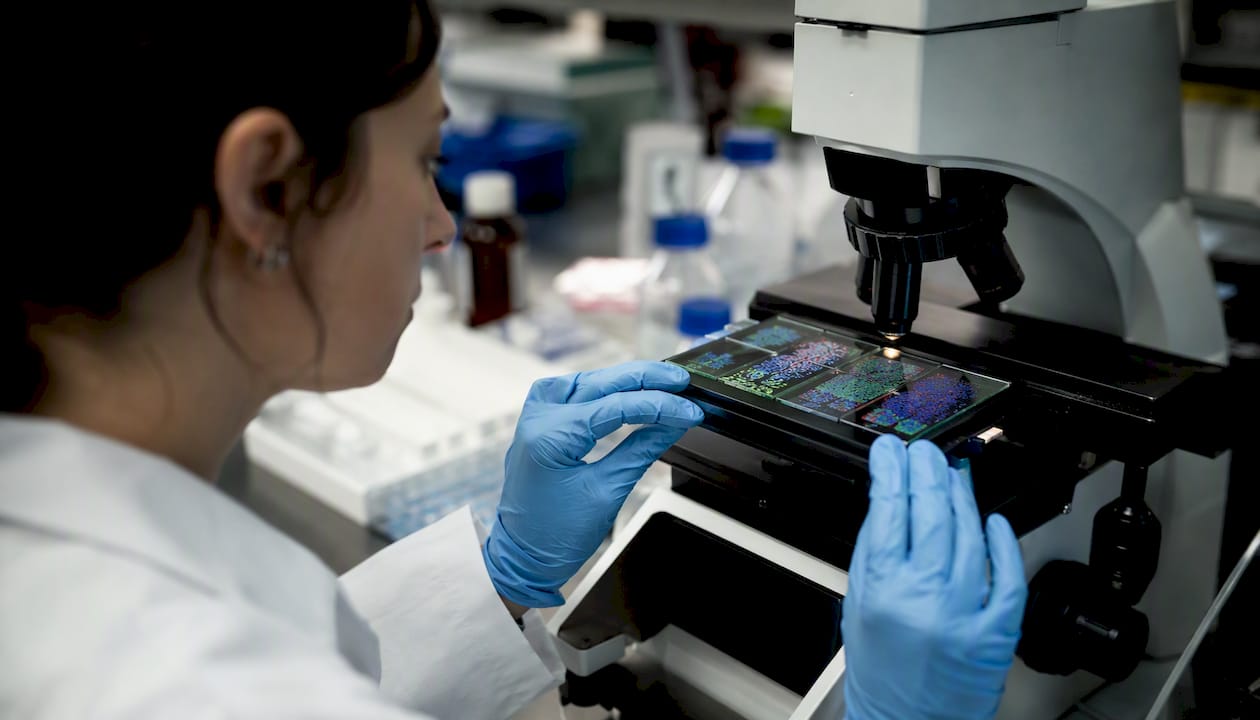
The tumor microenvironment (TME) is the local ecosystem of nonmalignant cells, matrix, and signals that actively controls tumor growth, immune evasion, metastasis, and therapy response. Understanding it is no longer optional for anyone working in oncology. It is the decisive terrain where cancers survive, adapt, and resist treatment.
- The TME drives resistance. Stromal and immune components actively suppress anti-tumor immunity and shield cancer cells from chemotherapy, targeted agents, and immunotherapy.
- Spatial and metabolic heterogeneity matter. Tumor cells in hypoxic, acidic, or desmoplastic zones behave differently from those in well-perfused regions, and a single biopsy rarely captures that complexity.
- Targeting TME components improves outcomes. Immune checkpoint inhibitors, anti-angiogenic agents, and stromal normalization strategies all work, at least in part, by reprogramming the microenvironment rather than killing tumor cells directly.
- The field is moving fast. Spatial transcriptomics, single-cell sequencing, and liquid biopsy are transforming how we map and monitor the TME in real time.
This article moves through the TME’s cellular and acellular components, its vascular and metabolic architecture, immune evasion mechanisms, therapy resistance, metastasis, experimental models, and emerging therapeutic strategies, closing with translational challenges and HCRF-supported research highlights.
Key Takeaways
The tumor microenvironment actively controls cancer growth, immune evasion, metastasis, and therapy resistance, making it the central target for the next generation of durable oncology treatments.
| Point | Details |
|---|---|
| TME drives resistance | Stromal cells, ECM barriers, and metabolic suppression protect tumor cells from chemotherapy, targeted agents, and immunotherapy. |
| Spatial heterogeneity changes everything | Spatial ecotypes and single-cell profiling reveal immune-rich and immune-desert regions that bulk sequencing misses, altering prognosis and treatment selection. |
| Immune evasion is active, not passive | The TME recruits Tregs and MDSCs, expresses PD-L1, and depletes nutrients to silence anti-tumor immunity, requiring combinatorial reprogramming strategies. |
| ECM remodeling blocks delivery | Desmoplasia raises interstitial pressure and excludes immune cells; normalizing matrix mechanics is a legitimate therapeutic target, not just a delivery optimization. |
| Longitudinal sampling is essential | Single biopsies misrepresent TME composition; serial tissue and liquid biopsy are necessary to track adaptive resistance and guide treatment decisions. |
Table of Contents
- What makes up the tumor microenvironment?
- How the extracellular matrix reshapes the tumor’s physical world
- Why tumor blood vessels work against treatment
- Metabolic competition inside the tumor
- Heterogeneity, plasticity, and the problem of a moving target
- How the TME silences the immune system
- How the TME drives therapy resistance and recurrence
- The TME’s role in metastasis and pre-metastatic niche formation
- Which experimental models best capture TME complexity?
- Therapeutic strategies that target the tumor microenvironment
- Translational barriers and the research priorities that matter most
- HCRF-supported research: from mechanism to the clinic
- Why TME research is the most urgent frontier in oncology right now
- Sources
What makes up the tumor microenvironment?
The TME is not a passive backdrop. It is an active, bidirectional ecosystem in which tumor cells and their neighbors continuously reshape one another. Two broad compartments define it: immune cells and stromal cells.
Immune cells
Understanding tumor cell origins helps contextualize why immune responses vary so dramatically across cancer types.
- CD8+ cytotoxic T cells: The primary anti-tumor effectors. Their density and functional state within a tumor correlate with prognosis across many solid cancers. The TME often exhausts them through chronic antigen exposure and inhibitory signals.
- CD4+ helper T cells and regulatory T cells (Tregs): Helper T cells amplify immune responses; Tregs suppress them. Tumors actively recruit Tregs via CCL22 and TGF-β, tipping the balance toward tolerance.
- B cells: Context-dependent. Tertiary lymphoid structures rich in B cells associate with better immunotherapy responses in several tumor types, yet some B cell subsets can suppress immunity.
- Natural killer (NK) cells: Innate killers that recognize stress ligands on tumor cells without prior sensitization. The TME downregulates NK-activating receptors and reduces their cytotoxic capacity.
- Dendritic cells (DCs): Antigen-presenting cells that bridge innate and adaptive immunity. Dysfunctional or tolerogenic DCs in the TME impair T cell priming and sustain immune ignorance.
- Neutrophils: Polarize into anti-tumor (N1) or pro-tumor (N2) phenotypes depending on TME signals. N2 neutrophils promote angiogenesis and metastasis.
- Myeloid-derived suppressor cells (MDSCs): Immature myeloid cells that accumulate in tumors and potently suppress T cell and NK cell function through arginase-1, reactive oxygen species, and nitric oxide.
- Tumor-associated macrophages (TAMs): Among the most abundant immune cells in many solid tumors. M2-polarized TAMs secrete VEGF, IL-10, and TGF-β, promoting angiogenesis, immune suppression, and invasion. High TAM density correlates with poor prognosis in several solid cancers, including breast, ovarian, and lung types.
High TAM infiltration is one of the most consistent negative prognostic signals across solid tumors. Repolarizing TAMs from an M2-like to an M1-like state is an active therapeutic strategy, with CSF1R inhibitors and CD40 agonists among the approaches in clinical development.
Stromal cells
- Cancer-associated fibroblasts (CAFs): Activated fibroblasts that deposit collagen, secrete growth factors (HGF, FGF), and remodel the ECM. CAFs are not a single population; at least four functional subtypes have been described, with distinct pro- and anti-tumor roles.
- Endothelial cells: Line tumor blood vessels. Tumor-derived VEGF drives their proliferation into structurally abnormal, leaky vessels that paradoxically reduce perfusion and drug delivery.
- Pericytes: Wrap around vessel walls and regulate permeability. In tumors, pericyte coverage is sparse and irregular, contributing to vascular dysfunction and hypoxia.
Pro Tip: When designing co-culture experiments, include at least one CAF subtype and one macrophage population alongside tumor cells. Omitting stromal partners systematically underestimates drug resistance and overestimates immune killing in vitro.
How the extracellular matrix reshapes the tumor’s physical world
The extracellular matrix is the scaffold within which all TME cells live, and tumors remodel it aggressively. Desmoplasia, the pathological accumulation of dense fibrous matrix, creates diffusion barriers that block immune cell infiltration and impair drug delivery, making it one of the most underappreciated drivers of therapy resistance.
ECM components and remodeling mechanisms:
- Collagens (especially type I and IV): The most abundant structural proteins. Cross-linking by lysyl oxidase (LOX) stiffens the matrix and activates integrin-mediated pro-survival signaling in tumor cells.
- Laminins: Basement membrane glycoproteins that regulate cell polarity, adhesion, and migration. Altered laminin isoforms promote invasion.
- Proteoglycans (hyaluronan, versican, perlecan): Regulate water retention, growth factor sequestration, and cell signaling. Hyaluronan accumulation raises interstitial fluid pressure.
- Matrix metalloproteinases (MMPs): Zinc-dependent enzymes secreted by tumor cells, CAFs, and macrophages that degrade matrix barriers, release matrix-bound growth factors, and open invasion corridors.
Biomechanical consequences:
- Increased tissue stiffness activates mechanosensing pathways (YAP/TAZ, FAK) that promote proliferation and resistance to apoptosis.
- Elevated interstitial fluid pressure reduces convective drug transport, meaning many chemotherapeutics never reach therapeutic concentrations at the tumor core.
- Dense ECM physically excludes CD8+ T cells from the tumor parenchyma, creating the “immune-excluded” phenotype seen in pancreatic ductal adenocarcinoma and some breast cancers.
ECM stiffness is not just a structural problem — it is a signaling problem. Mechanotransduction through integrin-FAK-YAP axes converts physical rigidity into transcriptional programs that sustain stemness, drug efflux, and immune evasion. Targeting matrix mechanics is therefore a legitimate therapeutic entry point, not merely a delivery optimization.
Pro Tip: In preclinical drug delivery studies, measure tumor stiffness by atomic force microscopy or shear-wave elastography alongside drug penetration assays. Reporting drug concentration at the tumor rim versus core reveals delivery failures invisible in whole-tumor homogenate assays.
NCI-supported research priorities explicitly include ECM remodeling as a central driver of cancer progression and therapy resistance, underscoring its translational urgency.
Why tumor blood vessels work against treatment
Tumors cannot grow beyond a few millimeters without recruiting new blood vessels, yet the vasculature they build is structurally and functionally abnormal. This paradox sits at the heart of why many therapies fail to reach their targets.
Primary angiogenic mediators:
- VEGF-A: The dominant pro-angiogenic signal, upregulated by HIF-1α under hypoxia. Drives endothelial proliferation and vessel sprouting.
- ANGPT1/ANGPT2 and TIE2 signaling: ANGPT1 stabilizes vessels; ANGPT2, overexpressed in tumors, destabilizes them and promotes leakiness.
- HIF-driven pathways: Hypoxia-inducible factors transcriptionally activate VEGF, PDGF, and other angiogenic genes, creating a self-reinforcing hypoxia-angiogenesis loop.
- FGF, PDGF, and EGF: Secondary mediators that sustain vessel growth and recruit pericytes, though tumor pericyte coverage remains deficient.
Functional consequences of abnormal vasculature:
- Leaky, tortuous vessels create heterogeneous blood flow, leaving large tumor regions chronically hypoxic and acidic.
- Poor perfusion reduces the delivery of oxygen-dependent cytotoxics and limits the trafficking of immune effector cells.
- Abnormal pericyte coverage increases vessel permeability, contributing to elevated interstitial pressure.
Anti-angiogenic therapy (VEGF/VEGFR inhibition) was initially expected to starve tumors of blood supply. The clinical reality is more nuanced: low doses can transiently “normalize” vessels, improving perfusion and drug delivery, while high doses prune vessels and worsen hypoxia. Timing normalization with immunotherapy or chemotherapy is a critical design question in current trials.
Lymphangiogenesis, driven by VEGF-C and VEGF-D acting on VEGFR-3, creates new lymphatic channels that tumor cells exploit for regional dissemination. Sentinel lymph node involvement remains a key staging marker and a direct readout of lymphatic TME remodeling.
Metabolic competition inside the tumor
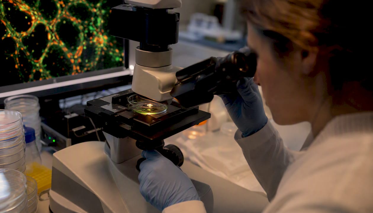
Cancer cells reprogram their metabolism, and the consequences extend far beyond the tumor cell itself. The metabolic TME is a battleground where tumor cells, stromal cells, and immune cells compete for the same limited nutrients.
Core metabolic shifts:
- Warburg effect: Even in the presence of oxygen, tumor cells preferentially use aerobic glycolysis, generating ATP rapidly but inefficiently and producing large amounts of lactate.
- HIF-1α and HIF-2α: Master transcription factors activated by hypoxia that upregulate glucose transporters (GLUT1, GLUT3), glycolytic enzymes, and VEGF. They also suppress oxidative phosphorylation.
- Lactate accumulation: Exported via monocarboxylate transporters (MCT1, MCT4), lactate acidifies the TME (pH often 6.5–6.9), which inhibits T cell cytokine production and NK cell killing.
- Nutrient competition: Tumor cells consume glucose and glutamine at rates that starve infiltrating T cells, impairing their proliferation and effector function.
Tumor-stroma metabolic cooperation:
CAFs can engage in “reverse Warburg” metabolism, producing lactate and pyruvate that fuel oxidative phosphorylation in adjacent tumor cells. Adipocytes in the TME supply fatty acids that support tumor cell survival under nutrient stress.
Metabolic targets under investigation:
- IDO1/IDO2 (indoleamine 2,3-dioxygenase): Depletes tryptophan and generates kynurenines that suppress T cells and expand Tregs.
- Glutaminase (GLS): Blocking glutamine catabolism starves tumor cells and may restore T cell function.
- MCT1/MCT4 inhibitors: Trap lactate inside tumor cells, inducing metabolic stress.
- Arginase-1: Expressed by MDSCs and TAMs, depletes arginine needed for T cell receptor signaling.
Metabolic suppression is one reason why immunotherapy fails in metabolically hostile tumors. Restoring T cell fitness by correcting the nutrient and pH environment is an active research priority, and metabolic and epigenetic reprogramming represent promising axes for both therapy and biomarker development.
Heterogeneity, plasticity, and the problem of a moving target
No two regions of a tumor are identical, and no tumor stays the same over time. This is the central challenge of TME biology: you are always studying a snapshot of something that is continuously evolving.
Intratumoral heterogeneity:
- Genetic heterogeneity: Subclones with distinct driver mutations coexist within a single tumor, each potentially interacting differently with the TME.
- Epigenetic heterogeneity: Chromatin states and DNA methylation patterns vary across cells with identical genomes, driving divergent gene expression programs.
- Phenotypic heterogeneity: Even genetically identical cells adopt different functional states depending on their local niche, oxygen tension, and signaling inputs.
- Spatial ecotypes: Spatial profiling tools now map how cells are physically organized within tumors, revealing that immune-rich and immune-desert regions can coexist within millimeters of each other. These spatial ecotypes carry prognostic and predictive information that bulk sequencing misses entirely.
Plasticity mechanisms:
- Epithelial-to-mesenchymal transition (EMT): Tumor cells shed epithelial markers and acquire mesenchymal properties, gaining invasive capacity and therapy resistance. EMT is reversible and context-dependent.
- Therapy-induced phenotypic shifts: Chemotherapy and targeted agents can select for or induce stem-like, drug-tolerant persister cells that repopulate the tumor after treatment.
- Transdifferentiation: Rare but documented; tumor cells can adopt lineage identities distinct from their cell of origin, confounding targeted therapy.
Senescence and aging stroma:
Senescent fibroblasts secrete a pro-inflammatory cocktail called the senescence-associated secretory phenotype (SASP), which includes IL-6, IL-8, MMPs, and VEGF. SASP promotes tumor cell proliferation, immune evasion, and angiogenesis. Aging stroma accumulates senescent cells, which may partly explain why cancer incidence and therapy resistance increase with age.
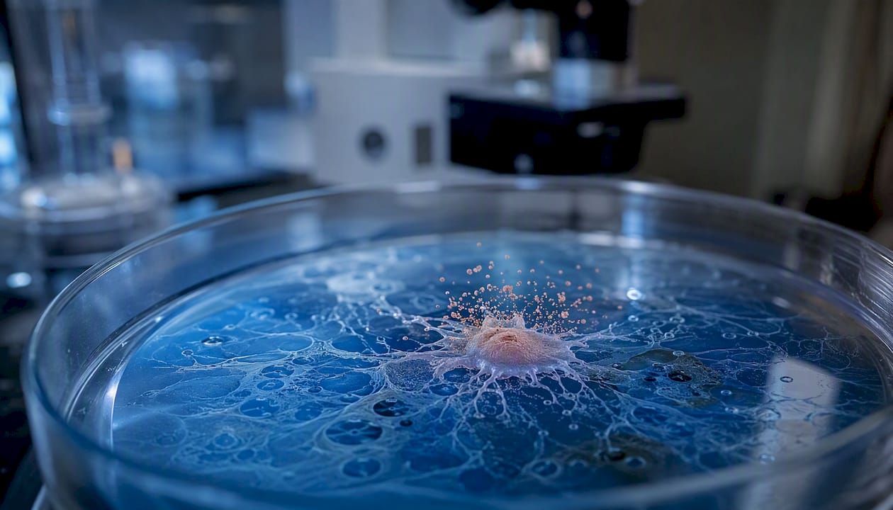
A single biopsy captures one spatial and temporal snapshot. Longitudinal sampling and liquid biopsy are not optional extras; they are methodological necessities for understanding TME evolution during treatment.
How the TME silences the immune system
Immune evasion is not a passive failure of immune surveillance. It is an active, TME-driven program, and durable responses to immunotherapy will require strategies that both kill tumor cells and reprogram the immunosuppressive elements around them.
Mechanism → effect → clinical consequence:
- PD-L1 expression on tumor and stromal cells → T cell exhaustion and anergy → reduced response to PD-1/PD-L1 checkpoint blockade
- Treg recruitment via CCL22/TGF-β → suppression of CD8+ T cell and NK cell activity → immune tolerance and tumor growth
- MDSC accumulation → arginase-1 and ROS-mediated T cell suppression → impaired cytotoxic responses
- TAM M2 polarization → IL-10 and TGF-β secretion → anti-inflammatory niche, angiogenesis, invasion
- Antigen presentation defects (MHC-I downregulation, beta-2 microglobulin loss) → T cells cannot recognize tumor cells → checkpoint inhibitor resistance
- ECM-mediated immune exclusion → physical barrier to T cell infiltration → “immune-excluded” or “immune-desert” phenotype
- Inhibitory cytokines (TGF-β, IL-10, IL-35) → broad suppression of DC maturation, NK activity, and T cell proliferation
The distinction between ‘hot’ and ‘cold’ tumors is not binary — it is a spectrum shaped by the TME. Hot tumors (inflamed, T cell-rich) respond better to checkpoint blockade, while cold tumors (excluded or desert) require strategies that first remodel the TME before immune killing can occur. Identifying which barrier dominates in a given patient’s tumor is the key clinical question.
The TME enforces immune suppression through active recruitment of suppressor cells, expression of inhibitory ligands, metabolic constraints, and inhibitory cytokines, all of which reduce checkpoint inhibitor effectiveness in cold tumors. Immune evasion is an active, TME-driven program, and durable responses require combinatorial strategies that reprogram immunosuppressive TME elements.
How the TME drives therapy resistance and recurrence
Therapy resistance is rarely just about the tumor cell. The surrounding microenvironment provides physical shelter, metabolic support, and signaling inputs that help cancer cells survive treatment and repopulate after apparent remission.
Mechanisms of TME-driven resistance:
- Physical barriers: Dense ECM and elevated interstitial pressure prevent adequate drug concentrations from reaching tumor cells, particularly in pancreatic and desmoplastic breast cancers.
- Drug sequestration: CAFs and TAMs can absorb and metabolize chemotherapeutics, reducing the effective dose reaching tumor cells.
- Niche-protected cancer stem cells: Perivascular and hypoxic niches shelter stem-like tumor cells that express drug efflux pumps and resist apoptosis. These cells repopulate the tumor after cytotoxic treatment.
- Reactive stroma: Treatment itself can activate CAFs and macrophages, triggering a wound-healing response that paradoxically promotes tumor regrowth and metastasis.
- Stromal signatures and chemo response: Stromal gene expression signatures have been linked to poor chemotherapy response in breast and colorectal cancers, and are increasingly incorporated into clinical trial stratification.
Counter-strategies under investigation:
- Combination therapy pairing cytotoxics with stromal normalization agents (e.g., hyaluronidase, LOX inhibitors) to improve drug penetration.
- Metabolic modulators (IDO inhibitors, glutaminase blockers) to restore immune cell fitness alongside immunotherapy.
- Immune reprogramming: CSF1R inhibitors to deplete or repolarize TAMs, CD40 agonists to activate DCs and macrophages.
- Epigenetic drugs (HDAC inhibitors, DNMT inhibitors) to reverse therapy-induced phenotypic shifts and restore antigen presentation.
For clinicians: when a patient progresses after an initial response, consider whether the resistance pattern fits a TME-driven mechanism. Stromal-rich tumors on imaging, rising neutrophil-to-lymphocyte ratio, or loss of MHC-I expression on rebiopsy all point toward microenvironmental rather than purely cell-autonomous resistance. Spatial profiling and patient supplements during immunotherapy can both influence treatment outcomes in ways that are easy to overlook.
A 2026 Frontiers review identifies the TME as the central orchestrator of therapeutic resistance and highlights reprogramming strategies including nanotechnologies, metabolic modulators, and stromal normalization.
The TME’s role in metastasis and pre-metastatic niche formation
Metastasis is not simply a tumor cell escaping into the bloodstream. The primary tumor actively conditions distant organs to receive disseminated cells, and the TME is the architect of that preparation.
The metastatic cascade, TME-driven:
- Local invasion: MMPs secreted by CAFs and tumor cells degrade basement membrane. CAF-generated “invasion corridors” of aligned collagen fibers guide tumor cell migration.
- Intravasation: Macrophages at the tumor-vessel interface (TMEM doorways) facilitate tumor cell entry into blood vessels by secreting EGF and VEGF.
- Circulation survival: Tumor cells cluster with platelets and neutrophils in the bloodstream, evading NK cell killing and shear stress.
- Arrest and extravasation: Tumor cells arrest at distant capillary beds and extravasate, guided by chemokine gradients (CXCL12/CXCR4 in bone marrow, CXCL1 in liver).
- Pre-metastatic niche conditioning: The primary tumor sends exosomes, cytokines, and bone marrow-derived cells ahead to remodel distant stroma before tumor cells arrive.
Mediators of pre-metastatic niche formation:
- Tumor-derived exosomes: Carry integrins, miRNAs, and proteins that reprogram stromal and immune cells at distant sites. Exosome integrin composition predicts organ-specific metastasis patterns.
- VEGF and S100 proteins (S100A8, S100A9): Recruit bone marrow-derived myeloid cells that suppress local immunity and deposit fibronectin, creating a permissive landing zone.
- Fibronectin: Deposited by resident fibroblasts in response to tumor signals; serves as an adhesion substrate for arriving bone marrow-derived cells and tumor cells.
ECM remodeling, stromal cells, and secreted mediators contribute directly to metastatic niche preparation, and TME-targeted strategies can alter metastatic progression. Biomarkers from the pre-metastatic niche, including circulating exosome profiles and S100 protein levels, are under investigation as early detection tools.
Which experimental models best capture TME complexity?
No single model captures the full TME, and choosing the wrong one is one of the most common sources of failed translation from bench to bedside.
Model comparison:
| Model | Main strengths | Main weaknesses | Best for |
|---|---|---|---|
| 2D co-culture | Simple, scalable, mechanistically tractable | No spatial architecture, no matrix, no flow | Rapid mechanistic screens, cytokine signaling |
| 3D organoids | Preserves tumor architecture, patient-derived options | Limited immune and stromal components | Drug sensitivity, epithelial biology |
| Patient-derived xenografts (PDX) | Human tumor genetics, stromal humanization possible | Murine immune system, expensive, slow | Drug response prediction, biomarker validation |
| Syngeneic mouse models | Intact immune system, genetically tractable | Mouse TME differs from human; limited antigen diversity | Immunotherapy mechanism, checkpoint biology |
| Organ-on-a-chip | Controlled flow, co-culture, real-time imaging | Low throughput, complex fabrication | Vascular biology, drug transport |
| Ex vivo tumor explants | Preserves native TME architecture briefly | Short viability window, limited perturbation | Short-term drug response, immune function |
Key molecular and spatial techniques:
- Single-cell RNA sequencing (scRNA-seq): Resolves transcriptional states of individual TME cells; reveals rare populations and cell-state transitions invisible in bulk data.
- Spatial transcriptomics (Visium, MERFISH, Xenium): Maps gene expression to tissue coordinates; identifies spatial ecotypes and cell-cell proximity relationships. Spatial and temporal mapping of TME ecotypes enables more predictive biomarkers than bulk signatures.
- Multiplex immunofluorescence (mIF) and imaging mass cytometry: Quantifies 30–50 protein markers simultaneously in tissue sections with spatial resolution.
- Mass cytometry (CyTOF): High-dimensional single-cell protein profiling of dissociated tumors; no spatial context but deep phenotyping.
- Liquid biopsy (cfDNA, circulating tumor cells, extracellular vesicles): Noninvasive monitoring of TME-derived signals. Liquid biopsy measures capturing tumor-derived extracellular vesicles and cfDNA can reflect shifts in TME activity and may enable monitoring when tissue biopsy is infeasible.
Pro Tip: Preserve spatial context from the moment of tissue collection. Snap-freeze or fix tissue within 30 minutes of resection, and allocate dedicated cores for spatial transcriptomics alongside standard FFPE blocks. Spatial data collected retrospectively from suboptimally preserved tissue loses most of its interpretive value.
Therapeutic strategies that target the tumor microenvironment
The TME is no longer just a target of opportunity. It is a primary therapeutic front, and the strategies aimed at it span from approved agents to early-phase trials.
Therapy classes, mechanisms, and development stage:
-
Immune checkpoint inhibitors (ICIs): Block inhibitory receptors (PD-1, PD-L1, CTLA-4, LAG-3, TIM-3) to restore T cell activity. Approved across multiple solid tumors; response rates vary widely by TME phenotype.
-
CAR-T and adoptive cell therapies: Engineer or expand tumor-reactive T cells ex vivo and reinfuse them. Approved in hematologic malignancies; solid tumor applications face TME-driven trafficking and exhaustion barriers.
-
Oncolytic viruses: Selectively replicate in tumor cells, inducing immunogenic cell death and TME inflammation. Early-phase trials in melanoma, glioblastoma, and pancreatic cancer.
-
CAF-targeting agents: Deplete or reprogram CAFs using FAP-targeted therapies, TGF-β inhibitors, or CXCR4 antagonists. Preclinical to early-phase; combination with ICIs is the primary rationale.
-
Anti-angiogenics and vascular normalization: VEGF/VEGFR inhibitors reduce aberrant vasculature. Approved in renal cell carcinoma, hepatocellular carcinoma, and others. Normalization dosing strategies aim to improve perfusion and immune infiltration before pruning.
-
Metabolic modulators: IDO1 inhibitors, glutaminase inhibitors, and MCT inhibitors aim to restore immune cell fitness in nutrient-depleted TMEs. Phase I/II trials ongoing; IDO1 inhibitor combinations with ICIs have shown mixed results, highlighting the need for better patient selection.
-
Epigenetic drugs: HDAC inhibitors and DNMT inhibitors can upregulate MHC-I expression, restore antigen presentation, and sensitize tumors to immunotherapy. Early-phase combinations with ICIs are active.
-
Nanoparticle delivery systems: Lipid nanoparticles, polymeric nanoparticles, and antibody-drug conjugates can be engineered to penetrate dense ECM, target specific TME cell populations, or release payloads in response to TME stimuli (low pH, MMP activity). Preclinical to early-phase.
-
Microbiome-based interventions: Gut microbiome composition influences systemic immune tone and ICI response. Fecal microbiota transplantation and defined bacterial consortia are in early clinical trials for ICI-refractory melanoma and other cancers.
-
Stromal normalization agents: Hyaluronidase (pegvorhyaluronidase alfa) degrades hyaluronan to reduce interstitial pressure and improve drug delivery. Tested in pancreatic cancer combinations.
Combination design and sequencing: The most important open question is not which TME target to hit, but in what order and at what dose. Stromal normalization and vascular normalization can transiently improve drug delivery and immune infiltration, but timing and sequencing with immunotherapy are critical design questions. Biomarker-driven enrollment, using spatial TME phenotype, immune cell density, or liquid biopsy signatures, is increasingly standard in well-designed combination trials. For clinicians evaluating patients for pancreatic cancer immunotherapy trials, TME phenotype assessment is becoming a practical eligibility consideration.
Translational barriers and the research priorities that matter most
The science of the TME has outpaced our ability to translate it. The gap between mechanistic insight and durable clinical benefit is real, and closing it requires confronting specific, named barriers.
Top translational barriers:
- Intratumoral heterogeneity: No single TME target is uniformly expressed. Spatial and clonal heterogeneity means that therapies effective in one tumor region may be irrelevant in another.
- Adaptive resistance: Tumors rewire their TME in response to therapy. Depleting one suppressive cell type often triggers compensatory upregulation of another.
- Lack of validated predictive biomarkers: PD-L1 expression and tumor mutational burden are imperfect predictors. Spatial TME phenotype and ecotype-based signatures are more informative but not yet standardized.
- Delivery challenges: Dense ECM, elevated interstitial pressure, and abnormal vasculature limit the reach of most therapeutic modalities, including nanoparticles.
- Combination toxicity: Targeting multiple TME components simultaneously risks overlapping immune-related adverse events and dose-limiting toxicities.
Pro Tip: Build longitudinal sampling into trial design from the start. Pre-treatment, on-treatment (cycle 2–3), and progression biopsies, paired with serial liquid biopsy, allow you to track TME evolution and distinguish adaptive resistance from primary non-response. Adaptive trial designs that use these data to modify treatment arms in real time are the most efficient path forward.
Prioritized research agenda:
- Develop and validate spatial multi-omic biomarkers that predict response across tumor types.
- Design rational combination trials with pre-specified TME endpoints and adaptive arms.
- Build delivery platforms that overcome ECM and vascular barriers without systemic toxicity.
- Harmonize multi-site cohort data through shared ontologies and open data-sharing agreements. Fragmented datasets slow validation; consortia like TCGA and HTAN have demonstrated what coordinated data sharing can achieve.
Biopharma innovation trends, including AI-driven spatial analysis and next-generation delivery platforms, are accelerating the pace at which translational pathways move from mechanism to trial-readiness.
HCRF-supported research: from mechanism to the clinic
The Hippocratic Cancer Research Foundation funds “out of the box” research at the Robert H. Lurie Comprehensive Cancer Center of Northwestern University, with a deliberate focus on projects that connect TME biology to translational opportunity. Three areas of active support illustrate that commitment.
Immune reprogramming in desmoplastic tumors. One supported project investigates strategies to convert immune-excluded, CAF-rich tumors into inflamed phenotypes amenable to checkpoint blockade. The approach combines ECM-modulating agents with immune activators, addressing the physical and biochemical barriers described earlier in this article. Translational aim: identify a combination regimen and companion biomarker suitable for a phase I/II trial at the Lurie Cancer Center.
Spatial profiling of TME ecotypes. A second initiative applies multiplex immunofluorescence and spatial transcriptomics to patient tumor samples to map immune cell organization and identify ecotype signatures that predict ICI response. This work directly addresses the validated predictive biomarker gap identified as a top translational barrier above.
Metabolic modulation and T cell fitness. A third project examines how correcting the metabolic TME, specifically lactate acidification and nutrient depletion, restores CD8+ T cell function in solid tumors. The goal is to identify metabolic co-interventions that improve immunotherapy response rates in patients whose tumors are metabolically hostile.
We believe that the most important cancer breakthroughs of the next decade will come from researchers willing to ask uncomfortable questions about the tumor’s neighborhood, not just the tumor cell itself. HCRF exists to fund exactly that kind of courage.
Learn more about how your support drives this work at the Hippocratic Cancer Research Foundation, and explore how to donate to cancer research and drive real impact. For researchers and patient advocates interested in cross-disciplinary models and rare disease research infrastructure, rare disease nonprofit resources offer additional context on how mission-driven organizations accelerate translational science.
Why TME research is the most urgent frontier in oncology right now
We are at an inflection point. The tools to map the TME with single-cell and spatial resolution now exist. The therapeutic strategies to reprogram it are entering trials. What is missing is the sustained, courageous funding that lets researchers follow the science wherever it leads, even when the path is unconventional.
At HCRF, we believe that understanding the tumor’s ecosystem is not a supporting act to finding a cure. It IS the path to the cure. Every donor who supports our mission at the Hippocratic Cancer Research Foundation is investing in research that treats cancer as the complex, adaptive, community-level disease it truly is.
For researchers seeking collaboration or funding: reach out through the HCRF website to explore partnership opportunities with the Lurie Cancer Center team.
For clinicians looking for trial options: the pancreatic cancer immunotherapy guide is a practical starting point for TME-informed trial selection.
For patients and families: HCRF provides access to information, clinical trial resources, and a community that shares your determination. You are not alone in this fight, and the science is moving faster than ever.

Sources
- Definition of tumor microenvironment - NCI
- Tumor Microenvironment - PMC - NIH
- Nature article on spatial ecotypes (2026)
- Tumor microenvironment and cancer metastasis: molecular mechanisms and therapeutic implications — Frontiers (2024)
This article is general information, not a substitute for advice from a qualified doctor. Consult a qualified healthcare professional about your own circumstances before acting on anything here.
Recommended
- Hippocratic Cancer Research Foundation: Innovative Therapies | Tumor Cell Types: Cell Origins and Clinical Implications
- Hippocratic Cancer Research Foundation: Innovative Therapies | Types of Tumors in the Human Body: A Complete Guide
- Hippocratic Cancer Research Foundation: Innovative Therapies | Does Tumor Size Determine Cancer Stage? What You Need to Know
- Hippocratic Cancer Research Foundation: Innovative Therapies | 4 Types of Malignant Tumors: What You Need to Know


Shaina and Christos Lardakis are proud to serve as chairs for this year’s Wings to Cure Gala, supporting the Hippocratic Cancer Research Foundation. As the owner of Kanela, Christos believes in the importance of giving back to the community, and together with Shaina, they are dedicated advocates for cancer research.
Read More →