


Types of Tumors in the Human Body: A Complete Guide
July 24, 2026
Types of Tumors in the Human Body: A Complete Guide

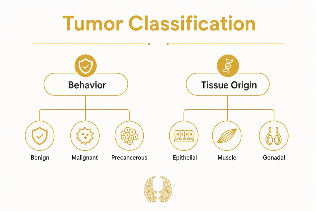
A tumor is an abnormal mass of tissue, and the most important thing to understand upfront is that “tumor” does not automatically mean cancer. The Cleveland Clinic classifies tumors into three core categories based on cellular behavior: benign, malignant, and precancerous. Each behaves differently, carries different risks, and demands a different clinical response.
Here is what separates them at a glance:
- Benign tumors grow locally, do not invade surrounding tissue, and do not spread to distant organs. They are noncancerous.
- Malignant tumors are cancerous. They invade neighboring tissue and can metastasize through the bloodstream or lymphatic system.
- Precancerous tumors contain abnormal cells that have not yet become malignant but will progress to cancer if left untreated or unmonitored.
- Tumors vs. cysts: A cyst is a fluid-filled sac. A tumor is a solid or semi-solid mass of abnormal cells. The two are not the same, though both can appear on imaging.
Understanding which category a tumor falls into is the foundation of every treatment decision that follows.
Table of Contents
- How tumors are classified by tissue origin
- How tumor behavior shapes clinical risk
- How tumors are diagnosed and managed
- What molecular biology reveals about tumor origins
- Why precancerous lesions deserve your full attention
- Key Takeaways
How tumors are classified by tissue origin
The tissue or cell type where a tumor originates determines its name, its behavior, and often its treatment. The National Cancer Institute organizes cancers into five primary categories based on cell of origin: carcinomas, sarcomas, leukemias, lymphomas and myelomas, and central nervous system cancers. Benign tumors follow the same tissue-origin logic.
Epithelial tumors (carcinomas)
Carcinomas arise from epithelial cells, the tissue that lines organs and body surfaces. They are the most common cancer type, affecting organs such as the breast, colon, skin, and bladder.
- Adenocarcinoma: forms in glandular epithelial cells; common in the breast, colon, and lung
- Basal cell carcinoma: the most frequently diagnosed skin cancer, arising from basal cells in the skin’s outer layer
- Squamous cell carcinoma: develops in flat squamous cells lining the skin, throat, and lungs
- Transitional cell carcinoma: originates in the bladder’s transitional epithelium
- Benign counterparts: adenomas (glandular tissue), papillomas (surface epithelium), and seborrheic keratoses
Connective and supportive tissue tumors (sarcomas)
Sarcomas develop from connective tissues including bone, cartilage, fat, muscle, and blood vessels. They are less common than carcinomas but often more aggressive.
- Osteosarcoma: malignant bone tumor, most common in adolescents
- Chondrosarcoma: arises from cartilage cells
- Rhabdomyosarcoma: originates in striated muscle
- Leiomyosarcoma: develops from smooth muscle
- Benign counterparts: fibromas (fibrous tissue), lipomas (fat), leiomyomas (smooth muscle, commonly called fibroids), and osteomas (bone)
Blood and lymphoid tumors
These tumors originate in hematopoietic and lymphoid cells. Leukemias arise from blood-forming cells in the bone marrow, while lymphomas develop in lymphoid tissue.
- Leukemias: include acute lymphoblastic leukemia (ALL), acute myeloid leukemia (AML), and chronic lymphocytic leukemia (CLL)
- Lymphomas: Hodgkin lymphoma and Non-Hodgkin lymphoma
- Multiple myeloma: a plasma cell malignancy
- Benign counterparts: plasmacytosis and certain myeloproliferative disorders that precede malignancy
Neural tumors
Neural tumors arise from cells of the brain, spinal cord, and peripheral nervous system.
- Gliomas and astrocytomas: originate in glial cells of the brain
- Meningiomas: arise from the meninges; usually benign but clinically significant due to location
- Schwannomas: develop from Schwann cells of peripheral nerves; typically benign
- Medulloblastoma: a malignant brain tumor more common in children
Muscle tumors
Smooth and striated muscle each give rise to distinct tumor types. Leiomyomas of the uterus are among the most common benign tumors in women. Their malignant counterpart, leiomyosarcoma, is rare but aggressive.

Gonadal tumors
Gonadal tumors originate in the ovaries or testes and include a uniquely complex group called germ cell tumors. These arise from multi-potential cells and can contain multiple tissue types within a single tumor. Examples include seminoma, choriocarcinoma, and teratoma. Stromal tumors of the gonads, such as Sertoli-Leydig cell tumors, arise from connective tissue and carry their own distinct naming conventions.
How tumor behavior shapes clinical risk
Growth rate and invasiveness separate benign from malignant tumors more reliably than any physical characteristic. Malignant tumors invade the basement membrane and spread; benign tumors do not. But the clinical picture is more nuanced than that binary suggests.
Physical exam findings alone cannot diagnose a tumor. Not all malignant tumors are hard or fixed in place, and not all benign tumors are soft or freely movable. A clinician who relies on feel rather than tissue analysis risks a serious misclassification.
Precancerous lesions occupy a middle ground that demands respect. They are not yet cancer, but they carry real progression risk. Colon polyps are a clear example: benign but capable of becoming malignant without surveillance and removal.
Location can make a benign tumor every bit as dangerous as a malignant one. A meningioma pressing on the brain can raise intracranial pressure to life-threatening levels, even though it carries no malignant cells. The tumor’s impact on function, not just its pathology label, drives urgency.
Pro Tip: Never assume a “benign” diagnosis means “no action needed.” Ask your oncologist specifically about the tumor’s location, growth rate, and functional impact before deciding on a watch-and-wait approach.
How tumors are diagnosed and managed
Diagnosis begins with imaging and clinical examination, but neither is definitive. Biopsy and microscopic pathology examination are the gold standard for distinguishing benign from malignant tumors. A surgeon cannot reliably determine tumor nature by feel during an operation alone.
Key steps in the diagnostic and management process:
- Imaging (MRI, CT, PET, ultrasound): identifies tumor location, size, and structural characteristics; guides biopsy planning
- Biopsy: removes a tissue sample for microscopic examination by a pathologist; the only definitive classification method
- Pathology report: determines cell type, grade, and whether the tumor is benign, malignant, or precancerous
- Staging (for malignant tumors): assesses how far cancer has spread; guides treatment intensity. Learn more about cancer staging and what each stage means clinically.
- Treatment options: surgery, radiation, chemotherapy, targeted therapy, and immunotherapy, selected based on tumor type, location, grade, and stage
- Surveillance (for precancerous and benign tumors): scheduled imaging or repeat biopsy to monitor for progression
Management is never one-size-fits-all. A small, slow-growing lipoma in a non-critical location may need no treatment at all. An aggressive adenocarcinoma of the colon may require surgery, chemotherapy, and long-term follow-up within weeks of diagnosis.
What molecular biology reveals about tumor origins
Every tumor begins with a change at the cellular level. Oncogenes, when mutated, push cells to divide uncontrollably. Tumor suppressor genes, when silenced or deleted, remove the brakes on that division. Understanding which molecular drivers are active in a specific tumor has transformed how oncologists classify and treat cancer.
Targeted therapies now exist precisely because of this molecular knowledge. A tumor’s tissue origin tells you what it is; its molecular profile tells you how to fight it. Matching patients to personalized clinical trials and targeted therapies depends on knowing both.
Infectious agents add another layer of complexity. Virus-associated tumors include cervical cancer linked to human papillomavirus (HPV), Burkitt lymphoma linked to Epstein-Barr virus, and Kaposi sarcoma linked to human herpesvirus 8 (HHV-8). These viral connections have opened doors to prevention strategies, most notably the HPV vaccine, which directly reduces cervical cancer risk.
Carcinomas, which arise from epithelial cells, account for the majority of cancer diagnoses in the United States. That prevalence is why so much cancer research focuses on epithelial tissue biology and why early detection programs target organs lined with epithelial cells, such as the colon, cervix, and breast.
The Hippocratic Cancer Research Foundation (HCRF) supports exactly this kind of forward-thinking science at the Robert H. Lurie Comprehensive Cancer Center of Northwestern University. By funding research that goes beyond conventional approaches, HCRF helps translate molecular discoveries into real treatments for real patients.
Why precancerous lesions deserve your full attention
Precancerous lesions are the body’s warning signal, and catching them early is one of the most powerful tools we have in cancer prevention. These lesions contain abnormal cells that have not yet crossed into malignancy, but the trajectory is clear without intervention.
Colon polyps are the most studied example. Adenomatous polyps can progress to colorectal cancer over years, which is exactly why colonoscopy guidelines recommend screening starting at age 45 for average-risk adults. Removing a polyp during a routine colonoscopy can prevent a cancer from ever forming.
Other common precancerous conditions include cervical dysplasia (detected through Pap smears and HPV testing), Barrett’s esophagus (a precursor to esophageal adenocarcinoma), actinic keratosis on sun-damaged skin, and leukoplakia in the oral cavity. Each carries its own surveillance schedule and intervention threshold.
Surveillance frequency depends on lesion type, size, and individual risk factors such as family history and prior cancer diagnoses. A gastroenterologist managing Barrett’s esophagus may recommend endoscopy every one to three years; a dermatologist monitoring actinic keratoses may treat them immediately with cryotherapy to prevent progression. The common thread is that watchful waiting without a clear plan is not the same as active surveillance. Understanding what causes cancer at the cellular level helps explain why these lesions progress and why early action matters so much.
Key Takeaways
Tumors are classified by cellular behavior and tissue origin, and that classification directly determines treatment. Biopsy remains the only definitive diagnostic method.
| Point | Details |
|---|---|
| Three core tumor categories | Benign, malignant, and precancerous tumors each carry distinct risks and require different clinical responses. |
| Tissue origin drives naming | Carcinomas arise from epithelial cells, sarcomas from connective tissue, and leukemias from blood-forming cells. |
| Biopsy is definitive | Physical examination and imaging cannot replace microscopic pathology for accurate tumor classification. |
| Benign does not always mean safe | Benign tumors in critical locations, such as meningiomas near the brain, can cause life-threatening complications. |
| Precancerous lesions need surveillance | Lesions like colon polyps require active monitoring and often removal to prevent progression to malignancy. |
With love and gratitude, we stand with every patient, every family, and every researcher carrying hope forward. The HCRF is here, and so are you.
Support the mission. Join us at HCRF and help fund the research that changes lives.

Recommended
- Hippocratic Cancer Research Foundation: Innovative Therapies | How Many Stages of Cancer Are There? A Clear Guide
- Hippocratic Cancer Research Foundation: Innovative Therapies | What Causes Cancer: Key Factors and Prevention
- Hippocratic Cancer Research Foundation: Innovative Therapies | Cancer Awareness Month Calendar: 2026 Complete Guide
- Hippocratic Cancer Research Foundation: Innovative Therapies | Supplements to Avoid During Immunotherapy: A Patient Guide
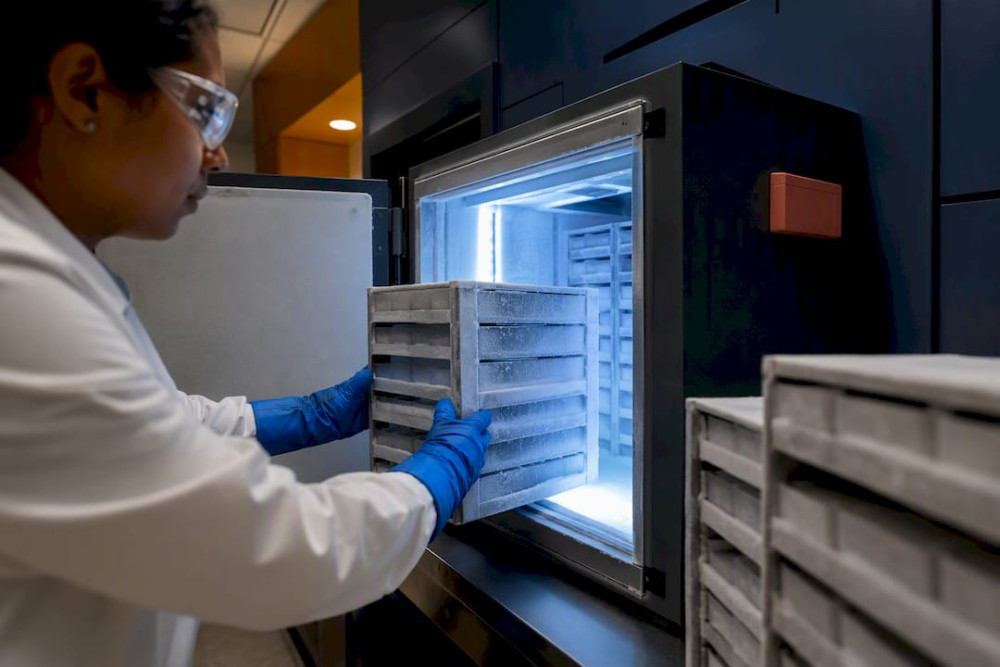
A donor guide to biobank tissue donation: what to expect, your consent and privacy rights, how samples are used, and where to register.
Read More →
Featured in Modern Luxury’s Dynamic Women 2026 for her philanthropic leadership and dedication to cancer research.
Read More →