

AI in Cancer Research for Researchers and Funders: Prioritize Data
September 3, 2026
AI in Cancer Research for Researchers and Funders: Prioritize Data
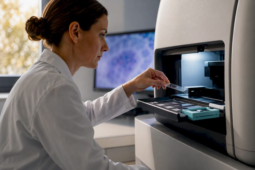
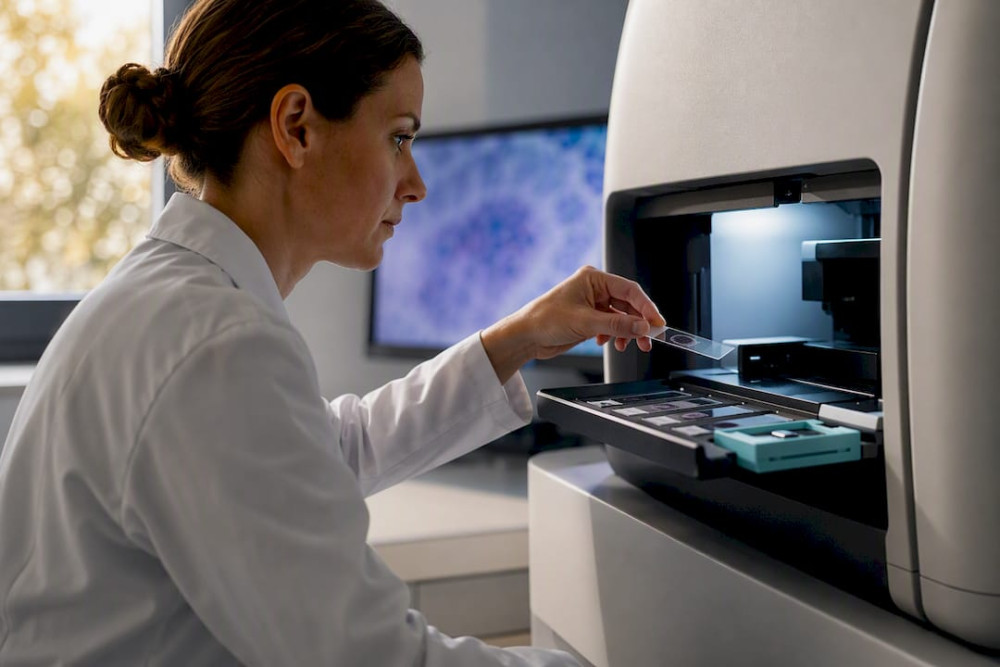
AI in cancer research already earns its place in two areas: imaging and pathology diagnostics, and drug discovery pipelines, where retrospective evidence is strong and growing. A multimodal breast cancer system recently reported an AUC of 0.973, and AI-driven virtual screening is reshaping early drug candidate identification. Treatment personalization and adaptive therapy remain genuinely promising but still need prospective trials, standardized reporting, and stronger governance before they change bedside decisions at scale.
TL;DR:
- Validating AI tools across multiple centers and diverse datasets is crucial, as models trained on single-site data often underperform in different clinical environments.
- High AUC scores in retrospective studies need prospective trials measuring real-world outcomes like biopsy reduction and faster diagnosis to confirm clinical usefulness.
- Biases from non-representative datasets and lab variations cause models to fail when deployed outside their training conditions, necessitating harmonization and fairness testing.
- Most AI advancements in imaging and pathology are already operational, but genomic and multimodal approaches still require further validation and integration.
- Funding should prioritize data infrastructure, harmonized protocols, and clinical validation efforts over algorithm development for sustained impact in cancer AI.
Table of Contents
- Where AI Is Actually Changing Cancer Care Today
- Reading the Evidence: What the Best Studies Actually Show
- Why Strong Lab Results Don’t Always Survive Contact With the Clinic
- Getting From Promising Results to Real Clinical Practice
- The Next Frontier: Generative Models, Digital Twins, and Adaptive Therapy
- Why Philanthropic Funding Is the Missing Piece in This Story
- Sources
Where AI Is Actually Changing Cancer Care Today
The gap between “AI can do this in a paper” and “AI does this in a clinic” is the single most important thing to understand before reading any headline about artificial intelligence cancer therapy. Some applications have crossed that gap. Most haven’t yet. Here’s the honest map, organized by how far each has traveled from bench to bedside.
Imaging is the most mature front. Mammography screening tools now flag subtle density patterns and microcalcifications that fatigued radiologists miss on a tenth read of the day. CT-based lung nodule triage systems sort scans by malignancy risk, letting radiologists prioritize the studies that actually need urgent eyes. Radiomics, the practice of extracting quantitative texture and shape features from medical images that the human eye can’t parse, has moved from a research curiosity to a standard input in several triage pipelines. The National Cancer Institute documents this shift across screening and diagnosis as one of the clearest wins in the field, while also flagging that most gains still come from retrospective datasets rather than head-to-head clinical trials.
Pathology is close behind. Whole slide imaging (WSI) classifiers digitize tissue biopsies and flag regions of concern before a pathologist even opens the case. This matters practically: it can cut the number of benign biopsies patients undergo, sparing them unnecessary procedures and the anxiety that comes with a “possible cancer” workup. Recent work on multi-cancer histopathology classification using DenseNet-style transfer learning architectures reports high validation accuracy across several tumor types, though single-site training data remains a recurring weakness researchers are working to fix.
Genomics and multi-omics integration is where things get harder, and more interesting. Variant interpretation, molecular subtyping, and models that fuse genomic, transcriptomic, and imaging data are advancing quickly, but the underlying biology is messier than a mammogram. A tumor’s genomic profile interacts with its microenvironment, the patient’s immune status, and prior treatment exposure in ways that single-modality models simply can’t capture. This is precisely why multimodal approaches, models trained on imaging, pathology, and genomic data simultaneously, are pulling ahead of single-input systems in peer-reviewed comparisons.
Precision therapy selection is the frontier everyone wants but almost nobody has fully proven. Predictive models that forecast treatment response, prognosticate survival, or match patients to open clinical trials based on their molecular profile represent the biggest promise in oncology AI. They also carry the biggest evidence gap. Most of these models are trained and tested on the same population they were built from, which tells you little about how they’ll perform on a patient from a different hospital system, a different ancestry group, or a different disease stage.
Surveillance and population health round out the picture. Natural language processing tools now extract structured cancer data from unstructured electronic health record (EHR) notes, a task that used to require armies of manual chart abstractors. The Surveillance, Epidemiology, and End Results (SEER) program has explored automation here, and population-level risk prediction models are starting to flag patients for earlier screening based on combined lifestyle, genetic, and demographic signals.
A few applications worth watching closely as this technology matures:
- Mammography and lung CT triage tools that reduce radiologist workload on low-risk scans
- WSI-based pathology classifiers that cut unnecessary biopsy rates
- Multimodal models combining radiology, pathology, and genomic inputs for diagnostic confidence
- NLP-driven EHR extraction that speeds cancer registry reporting
- Trial-matching algorithms that connect patients to relevant clinical trials faster than manual review
Machine learning cancer diagnosis tools are, in short, doing their best work where the input data is already structured and abundant, images and slides. The messier the data, the further AI still has to go.
Reading the Evidence: What the Best Studies Actually Show
Numbers like “AUC of 0.97” sound definitive. They’re not, at least not on their own. An AUC (area under the receiver operating characteristic curve) close to 1.0 means a model distinguishes cancer from non-cancer almost perfectly on the data it was tested against. Whether that holds up on a different hospital’s scanners, with a different patient mix, is an entirely separate question, and it’s the question that determines whether a study becomes a clinical tool or stays a journal article.
A useful benchmark to hold every AI oncology study against: a multimodal breast cancer system called BINDS, trained and validated across 27,048 participants from eight centers and seven public datasets, reported an AUC of 0.973 and a potential 32.4% reduction in unnecessary benign biopsies. That scale of external validation, multiple centers, multiple public datasets, is rare, and it’s exactly why this study stands out from single-site work reporting similarly impressive numbers on far smaller cohorts.
Why does multi-center validation matter so much? Because a model trained entirely on one hospital’s imaging equipment learns that equipment’s fingerprint along with the cancer signal. Swap in a different scanner brand, a different staining protocol, or a different patient demographic, and performance can quietly collapse even though the reported AUC never changes on paper. The BINDS result clears that bar in a way many single-center studies don’t.
A few other evidence patterns worth flagging when you read AI oncology research:
- High-performance imaging and pathology studies frequently report AUCs above 0.90, but check whether the cohort came from one institution or several. Single-center numbers, even strong ones, tend to shrink on external testing.
- Multi-cancer histopathology classifiers using architectures like DenseNet report high validation accuracy across several tumor types simultaneously, a genuinely impressive technical feat, though these results still largely reflect retrospective, not prospective, testing.
- Drug discovery studies claim meaningful acceleration in candidate identification through virtual screening and generative molecular design, and network-based drug repurposing is helping researchers prioritize which existing compounds might work against new targets. These are real efficiency gains at the computational stage. They still require full wet-lab validation, toxicity screening, and eventually human trials before any of it reaches a patient.
The caveat that applies across nearly all of this evidence: retrospective validation (testing a model on data it wasn’t trained on, but that was collected in the past) is not the same as prospective validation (testing a model’s real-time decisions on new patients as they arrive). Almost everything described above sits in the retrospective category. That’s not a failure. It’s simply the necessary first step before the harder, slower, more expensive prospective trials that regulators and clinicians actually need to see.
Why Strong Lab Results Don’t Always Survive Contact With the Clinic
A model that scores 0.97 in a paper and then underperforms in a different hospital isn’t lying about its original results. It’s revealing a mismatch between the conditions it was built for and the conditions it’s now facing. Understanding why that happens is essential for anyone funding, designing, or evaluating deep learning cancer treatment research.
Batch effects are the quiet killer. Different scanners, different staining chemistries, different lab pipelines all introduce systematic variation that has nothing to do with biology, but that a model can accidentally learn to rely on anyway. A technical review on explainability and harmonization identifies this as one of the most common hidden causes of model failure when systems move between institutions. Harmonization techniques, domain adaptation methods, and deliberate data augmentation during training all help close this gap, but they have to be built in from the start, not patched on after deployment.
Dataset representativeness compounds the problem. A model trained mostly on one demographic group, one geographic region, or one disease stage will carry that bias into every prediction it makes, often silently. Fairness testing, deliberately checking model performance across age, sex, race, and disease-stage subgroups, needs to be a standard reporting requirement, not an optional appendix.
Explainability is judged differently than most people assume. Clinicians don’t necessarily want a mathematically elegant saliency map highlighting which pixels drove a prediction. They want reasoning that maps onto biology they recognize. A review on AI in oncology notes that multimodal alignment, models that combine radiology and pathology signals the way a tumor board actually reasons through a case, tends to earn clinician trust more effectively than abstract heat maps ever do.
Four practical fixes deserve priority in any translational research agenda:
- Build harmonization and domain adaptation into training from day one, rather than treating cross-site generalization as an afterthought.
- Report subgroup performance by default, breaking down accuracy by demographic and clinical variables instead of a single headline number.
- Prioritize multimodal, biology-aligned explainability methods over abstract visualization techniques that clinicians struggle to act on.
- Adopt federated learning where data sharing is legally or logistically restricted, allowing models to train across institutions without moving sensitive patient data across borders.
Pro Tip: When evaluating any AI oncology study, check first whether the validation cohort came from the same institution as the training data. If the answer is yes, treat the reported performance as a ceiling, not a guarantee, of what the model will do elsewhere.
Operational risk doesn’t end at deployment, either. Models can drift over time as patient populations, imaging equipment, and clinical protocols change, meaning a system validated well in 2024 may quietly degrade by 2027 without continuous monitoring. Regulatory and privacy constraints add another layer, particularly around how patient data can be pooled across institutions, which is exactly why federated learning, training a shared model without centralizing raw patient data, has become one of the more realistic near-term answers to the dataset-diversity problem.
Getting From Promising Results to Real Clinical Practice
The distance between a strong retrospective study and an FDA-cleared, workflow-integrated clinical tool is longer than most published papers let on. Closing it requires deliberate study design, not just better algorithms.
Prospective trials need to measure what clinicians actually care about. A high AUC on archived scans is a necessary starting point, not an endpoint. The metrics that matter for real translation are things like reduced unnecessary biopsies, shortened time to diagnosis, fewer missed cancers on follow-up, and measurable time saved in radiologist or pathologist workflow. The NCI’s own guidance on AI in cancer research explicitly calls for prospective trials and explainability standards as prerequisites for real clinical adoption, not optional extras.
Reporting needs a minimum bar that most current studies don’t clear. A defensible AI oncology study today should include demographic breakdowns of its training and test cohorts, at least one external validation cohort from a different institution, and ideally pre-registration of the study’s primary endpoints before data collection begins. Absent any one of these, treat the headline performance number with real skepticism.
Regulatory pathways matter more than researchers sometimes appreciate. In the United States, AI-based diagnostic tools most often move through FDA clearance or authorization pathways designed for software as a medical device, which typically require evidence of both analytical validity (does it measure what it claims to measure) and clinical validity (does that measurement actually improve care). Post-market surveillance, ongoing monitoring after a tool reaches clinical use, is increasingly expected too, since a model’s real-world performance can shift as the patient population it sees evolves.
Deployment is its own discipline, separate from model development. A few things need to happen before any AI tool touches a real patient workflow:
- Integration with existing EHR systems so the tool fits inside, rather than disrupts, existing clinical documentation
- Real-time performance monitoring to catch drift before it affects patient care
- Multidisciplinary oversight boards that include clinicians, data scientists, ethicists, and patient representatives
- Clear escalation protocols for when the AI’s recommendation and the clinician’s judgment disagree
Programs supporting this kind of rigorous translational work, the unglamorous but essential middle step between a promising paper and a usable clinical tool, are exactly where philanthropic funding tends to matter most. HCRF’s coverage of innovative cancer research partnerships explores how this translation actually happens on the ground, including the multi-institution collaborations that make prospective validation possible in the first place.
The Next Frontier: Generative Models, Digital Twins, and Adaptive Therapy
If diagnostics and drug discovery are where AI in cancer research has already proven itself, the next wave, generative AI and personalized simulation, is where the field is placing its biggest bets for the years ahead.
Generative AI for molecule design and adaptive treatment regimens is moving from concept to early proof. Rather than simply screening existing compound libraries, generative models can propose entirely new molecular structures optimized for a specific target, then hand those candidates off for lab synthesis and testing. A perspective on generative AI in cancer treatment describes this as a genuine pathway toward dynamically tuned, personalized treatment regimens, while being equally clear that clinical validation, data curation, and the risk of AI “hallucinations”, confident but incorrect outputs, remain unresolved barriers before any of this reaches routine practice.
Patient-specific digital twins are the most ambitious idea in the pipeline. The concept: build a computational model of an individual patient’s tumor, detailed enough to simulate how it might respond to different treatment sequences before a single one is tried on the actual patient. Early pilot work is promising, but the data infrastructure required, longitudinal imaging, genomic sequencing, treatment response history, all tied together for a single patient, is substantial. This remains an active research frontier rather than a deployed tool, and it will likely stay that way for several years yet.
Large multimodal models and language models are quietly speeding up the research process itself. Beyond direct clinical applications, these tools are being used to synthesize massive volumes of oncology literature and extract patterns from EHR data at a scale no research team could manage manually, accelerating hypothesis generation even before a single new trial begins.
Federated learning remains the most realistic near-term enabler of all of this. Every one of these frontiers, generative drug design, digital twins, multimodal foundation models, needs large, diverse datasets to work safely. Federated learning lets institutions train shared models collaboratively without centralizing sensitive patient data in one place, which is likely to be the practical bridge between today’s single-institution models and tomorrow’s genuinely generalizable ones.
A short list of what to watch over the next several years:
- Generative molecule design paired with faster wet-lab validation cycles
- Early digital twin pilots focused on narrow, well-defined tumor types before broader rollout
- Multimodal foundation models trained jointly on imaging, pathology, and genomic data
- Federated learning consortiums spanning multiple cancer centers
Pro Tip: If you’re evaluating where to direct research funding or attention, prioritize programs building the underlying data infrastructure, harmonized imaging protocols, federated learning networks, over programs chasing the next flashy algorithm. The bottleneck in this field is data, not math.
Why Philanthropic Funding Is the Missing Piece in This Story
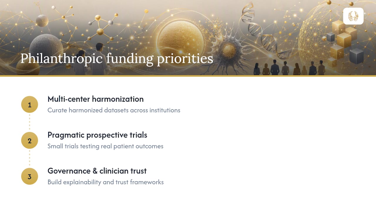
Here’s what most coverage of AI in cancer research leaves out: the algorithms are rarely the bottleneck anymore. The bottleneck is unglamorous, expensive, multi-year infrastructure work, curating harmonized datasets across institutions, running small pragmatic prospective trials, building explainability tools that clinicians actually trust. That work doesn’t attract venture capital the way a flashy new model does, and it doesn’t fit neatly into a single grant cycle. It’s exactly the kind of “out of the box” work HCRF exists to fund at the Robert H. Lurie Comprehensive Cancer Center of Northwestern University.
We believe the smartest philanthropic dollars in this space right now go toward three things: multi-center dataset harmonization projects that let researchers validate models beyond a single hospital’s walls, pragmatic small-scale prospective trials that test whether a promising retrospective result actually changes patient outcomes, and governance frameworks that build clinician trust before a tool ever reaches a bedside. None of these make headlines the way a 0.97 AUC does. All three are what separate a genuinely useful clinical tool from a permanently promising paper.
If you’re a researcher, this is the funding gap worth naming explicitly in your next grant application. If you’re a donor or corporate sponsor, this is where your contribution does the most durable good, not in funding one more algorithm, but in funding the validation infrastructure that lets good algorithms actually reach patients. Explore how Hcrfwingstocure directs support toward this kind of translational work, or see our donor’s guide to funding cancer research for specific ways to get involved.
The promise of precision oncology has never been in doubt. What’s been in doubt is whether the field builds the patient, unglamorous groundwork required to make that promise real. We intend to keep funding exactly that groundwork.
— HCRF
This article is general information, not a substitute for advice from a qualified doctor. Consult a qualified healthcare professional about your own circumstances before acting on anything here.
Sources
- Artificial intelligence for precision oncology from phenotyping and drug discovery to clinical translation | Discover Oncology
- Artificial Intelligence (AI) and Cancer | NCI
Recommended

Four checks patients and caregivers can use to read clinical trial results, find absolute benefit and NNT, spot bias, and know what to ask.
Read More →
Thank you for an unforgettable Cocktails for a Cure—relive the night in our photo gallery. See you at Wings to Cure this November!
Read More →