

3 Proven Uses of AI in Cancer Imaging for Clinicians and Funders
September 21, 2026
3 Proven Uses of AI in Cancer Imaging for Clinicians and Funders
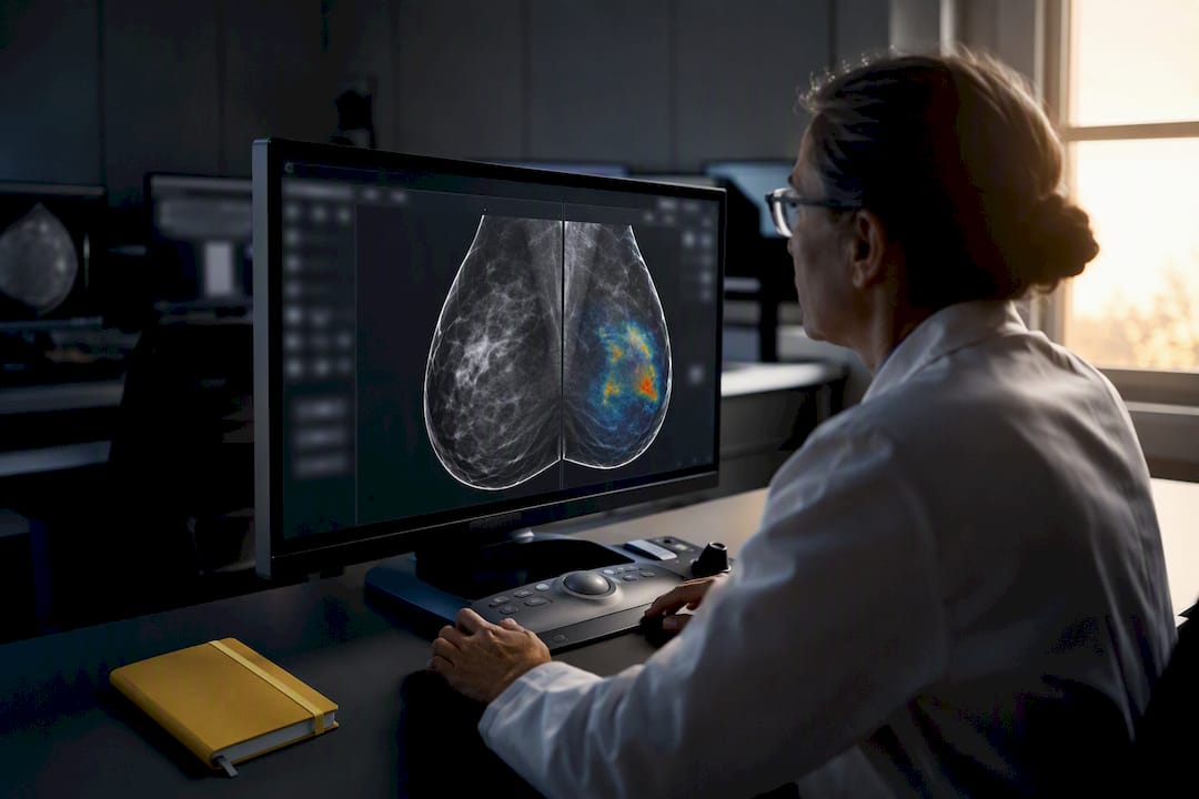
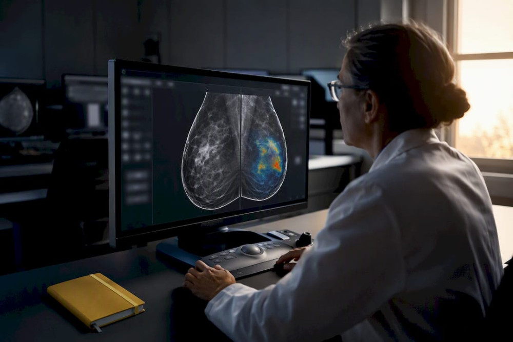
AI in cancer imaging now delivers proven gains in three domains: mammography screening, chest radiograph or CT triage, and digital pathology slide analysis. In each, the technology works as decision support, flagging cases and highlighting regions for a radiologist or pathologist, not as an autonomous diagnostician. That distinction rests on multicenter validation and prospective feasibility studies, and it comes with a catch every clinical team needs to plan for: post-deployment monitoring, because performance can shift once a model leaves its training environment.
TL;DR:
- AI in cancer imaging is mainly used as decision support, requiring continuous post-deployment monitoring due to performance shifts outside validation environments.
- Validation must include external, multicenter studies, with sensitivity, specificity, and localization accuracy offering more practical insight than AUC alone.
- Deployment strategies include triage, concurrent review, and automated reporting, each with specific workflow benefits and limitations.
- Major risks involve distribution shift, demographic bias, and limited explainability, which can be mitigated through ongoing monitoring and diverse data use.
- Reproducible research relies on open datasets like TCIA, MIMIC-CXR, and RSNA benchmarks, emphasizing pre-registration, shared code, and transparency for validation.
Table of Contents
- How Does AI Work in Cancer Imaging Technically?
- Where Does AI Improve Cancer Screening and Diagnosis?
- What Metrics Actually Prove an AI Tool Works?
- How Do You Integrate AI Into an Imaging Workflow?
- What Are the Biggest Risks of AI in Cancer Imaging?
- Which Datasets Support Reproducible Cancer Imaging Research?
- What Should AI Research Priorities Look Like Next?
- What Ethical Questions Does AI Raise in Cancer Imaging?
- How Does Explainable AI Build Clinical Trust in Oncology?
- What Do Real Clinical Studies Show About AI Performance?
- A Clinician’s Checklist for Adopting AI Tools
- Support Prospective Validation Studies With HCRF
- Sources
- FAQ
How Does AI Work in Cancer Imaging Technically?
Three families of models drive most clinical tools today. Convolutional neural networks (CNNs) remain the workhorse for pixel-level tasks: detecting a suspicious mass on a mammogram, segmenting a tumor volume on CT, or flagging a lung nodule on chest X-ray. They excel at spatial pattern recognition but need large, labeled training sets to generalize well.
Transformers and vision-language models (VLMs) handle a different problem: connecting what a scan shows to what a report says. These architectures power tasks like automated impression generation and structured reporting, where the model has to reason across an image and translate findings into clinical language. Large language models (LLMs) sit on top of this layer, standardizing report structure, drafting patient-facing summaries, and assisting with staging language, though the MDPI review on LLMs in cancer imaging flags hallucination risk and inconsistent performance on genuinely visual tasks as real constraints, not theoretical ones.
Self-supervised and foundation-model approaches represent the newer wave. Rather than training a narrow model on one labeled task, these systems pretrain on large, often unlabeled imaging archives, then adapt to specific applications with less task-specific data. Researchers building generalizable tools increasingly prioritize diverse pretraining sources precisely because narrow training data is what causes models to fail outside their original hospital system.
What lands on a clinician’s screen varies by task. Detection and segmentation models output probability scores, heatmaps or regions of interest, and pixel-level masks for tumor boundaries. Multimodal systems output drafted impressions, structured JSON fields for downstream systems, or full narrative summaries. Knowing which category a tool belongs to tell you what kind of validation evidence you should demand before trusting its output.
Where Does AI Improve Cancer Screening and Diagnosis?
Screening mammography has the deepest evidence base. Large multicenter retrospective and prospective feasibility studies found AI achieved higher sensitivity and noninferior specificity compared with human readers, alongside measurable increases in cancer detection rate in real screening workflows, according to research published in Nature Cancer. The same body of work found something equally important: performance drifted once models moved from validation cohorts into live deployment, which is why the study’s authors call for recalibrated detection thresholds rather than a single fixed cutoff.
Triage and prioritization is the second proven use case. In emergency and oncology imaging queues, AI models that flag likely-positive chest X-rays or CT scans let radiologists reorder their worklists so urgent findings get read first. A narrative review in BMC Artificial Intelligence reports AUCs for validated triage and detection systems ranging roughly from 0.84 to 0.96 depending on the task, and it frames the clinical benefit explicitly as workflow acceleration, not diagnostic replacement.
Segmentation and quantitative response assessment form a third application. Manual RECIST measurements are slow and prone to interobserver variability; automated volumetric segmentation can standardize tumor measurement across treatment cycles and flag subtle growth a human eye might miss between scans. This matters most in oncology trials, where consistent, reproducible tumor measurement directly affects whether a drug looks effective.
Digital pathology is the newest frontier with some of the most striking translational results. Models trained on whole-slide images can now predict molecular markers and even patient survival directly from tissue morphology, according to reporting from the Harvard Gazette on a new multimodal cancer AI tool. The caveat is generalizability. Pathology slides vary by scanner, staining protocol, and lab preparation far more than radiology images do, and models trained at one institution frequently underperform when applied to slides from another.
What Metrics Actually Prove an AI Tool Works?
AUC is the metric everyone cites and the one most likely to mislead when read alone. A model can post an AUC of 0.95 in a curated validation set and still miss cases that matter most in practice, because AUC averages performance across the entire probability threshold rather than showing what happens at the specific operating point a clinic will actually use. Sensitivity, specificity, and localization accuracy (how precisely a heatmap or bounding box overlaps the true lesion) tell you more about real-world behavior, and cancer detection rate is the metric that matters most for screening programs specifically.
External, multicenter validation is non-negotiable. A model validated only on the data it was tuned against tells you almost nothing about how it will perform on a different scanner fleet, patient population, or acquisition protocol. The Nature Cancer study on breast screening AI is instructive here precisely because it separated retrospective performance from prospective, real-world feasibility results, and the two did not match perfectly.
Reader studies, where radiologists interpret the same cases with and without AI assistance, reveal something metrics alone cannot: how clinicians actually use the tool. Some reader studies find radiologists override correct AI flags out of habit, while others find discordant AI and human reads, when routed to a second reader for arbitration, meaningfully reduce false positives, a pattern documented in the BMC Artificial Intelligence review. Distribution shift, the gradual mismatch between training data and live clinical data, is the single most common reason a well-validated model degrades after rollout, and it is the core justification for scheduled recalibration rather than a one-time validation checkpoint.
How Do You Integrate AI Into an Imaging Workflow?
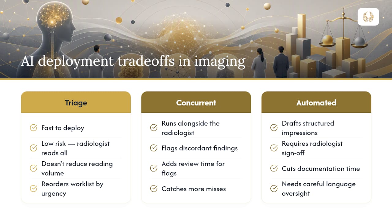
Three deployment patterns dominate current practice, and each carries distinct tradeoffs. Triage systems reorder worklists by predicted urgency; they’re fast to deploy and low-risk since a radiologist still reads every case, but they don’t reduce total reading volume. Concurrent or second-read systems run alongside the radiologist and flag discordant findings; they catch more misses but add review time for flagged discrepancies. Automated reporting systems draft structured impressions for radiologist sign-off, cutting documentation time but requiring careful oversight of language accuracy.
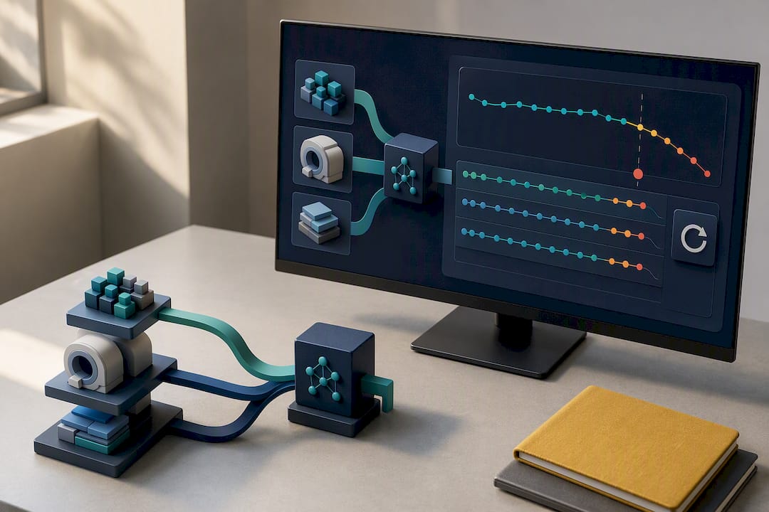
Technical integration means connecting the model to PACS and RIS systems, and increasingly the EHR, with attention to latency (a triage tool that takes ten minutes to score a scan defeats its own purpose) and to reporting templates that match your department’s existing structure.
Operationally, a workable pilot follows a clear sequence: run a limited pilot against retrospective and live cases, select and document your detection threshold explicitly, build a monitoring dashboard tracking subgroup performance and localization accuracy, and train staff on both the tool’s capabilities and its known failure modes. Governance matters as much as the technology. Threshold changes should be prespecified and approved, not adjusted informally when a radiologist feels the tool is “too sensitive” one week.
What Are the Biggest Risks of AI in Cancer Imaging?
Distribution shift tops the list. Models that perform well at validation can quietly degrade months into deployment as patient mix, scanner hardware, or acquisition protocols change, which is why continuous monitoring with per-subgroup dashboards and prespecified recalibration triggers has become standard practice in serious deployments, per findings from the Nature Cancer breast screening study.
Demographic bias is the related and harder problem. A model trained predominantly on one population’s imaging data can underperform for other groups, a risk that only shrinks with genuinely diverse training data, deliberate transfer learning to underrepresented populations, and routine fairness audits broken out by subgroup, not just aggregate accuracy.
Explainability remains partial. Saliency maps and heatmaps show where a model “looked,” not why it reached its conclusion, and newer agentic architectures that orchestrate multiple specialized models alongside a VLM to produce localized, step-by-step reasoning are showing promise for narrowing that gap, according to research on interpretable agentic AI systems published in npj Digital Medicine. Regulatory clearance and data governance round out the risk list: any tool touching identifiable patient imaging needs a clear data-handling policy well before FDA clearance status ever becomes the question that matters.
Which Datasets Support Reproducible Cancer Imaging Research?
Reproducibility in this field depends on a small set of open resources. The Cancer Imaging Archive (TCIA) is the standard source for annotated oncology imaging across modalities. MIMIC-CXR anchors chest radiograph research with paired reports. RSNA challenge datasets provide labeled benchmarks that let separate research teams compare methods on identical test sets.
Good practice means publishing your train/test/validation splits, sharing code alongside results, and pre-registering study design before results come in, since open evaluation on shared benchmarks is what lets other labs actually confirm a claimed result rather than take it on faith.
What Should AI Research Priorities Look Like Next?
The clearest translational bottleneck isn’t model performance, and resources like the RareLabs Knowledge Resource for Rare Disease Programs offer valuable support for research infrastructure and data curation needed to overcome it. It’s prospective, multi-site validation on genuinely diverse patient data, paired with explainability research and disciplined post-deployment monitoring. Nonprofit funding fills a specific gap here: it can back the unglamorous work, curated diverse datasets, arbitration studies, monitoring infrastructure, that traditional grant cycles often overlook in favor of flashier model development. Supporting innovative cancer research at a center with existing clinical trial infrastructure shortens the distance between a promising algorithm and a validated clinical tool. Clinician researchers evaluating a partnership should look for cancer centers that can link imaging AI work directly to active clinical trial pathways, since that link is what turns a retrospective finding into prospective proof.
What Ethical Questions Does AI Raise in Cancer Imaging?
Patient consent gets complicated the moment imaging data trains a model that will affect care decisions for people who never consented to that specific use. Most current consent frameworks were written for individual diagnostic use, not for a scan becoming training data for a system deployed years later at other institutions. Data ownership follows the same fault line: a hospital, a research consortium, and a technology vendor can each have a legitimate claim on a dataset, and unclear governance here has already stalled promising collaborations.
AI-induced disparity is the sharpest concern for clinical teams. A model trained mostly on data from academic medical centers serving one demographic profile can systematically underperform for patients outside that profile, and because the errors are statistical rather than obvious, they can persist for a long time before anyone notices the pattern. Mitigating this requires deliberately diverse training cohorts, subgroup-level performance audits published alongside aggregate accuracy, and a willingness to delay deployment when subgroup data is too thin to support a fairness claim.
None of this is solved by a single policy. It requires institutions and funders treating data diversity, consent transparency, and disparity auditing as core research deliverables, not afterthoughts addressed once a model already works well enough to publish. The NCI’s overview of AI in cancer research explicitly names data access and equitable implementation among the field’s central open challenges, and that framing is worth taking seriously rather than treating as boilerplate caveat language.
How Does Explainable AI Build Clinical Trust in Oncology?
Explainability in cancer imaging has moved past simple saliency maps, though those remain the most common technique. A saliency map highlights which pixels most influenced a model’s output, useful for a quick sanity check but limited because it shows correlation with the decision, not the reasoning behind it.
The more promising direction is agentic architecture. Systems like the interpretable framework described in npj Digital Medicine’s research on localized reasoning for radiology coordinate multiple specialized models and a vision-language layer to produce a step-by-step diagnostic narrative rather than a single opaque score. That structure lets a radiologist trace exactly which finding drove which conclusion, closer to how a human colleague would explain a read.
For LLM-generated reports specifically, explainability means something narrower but equally important: traceability back to the source finding. A drafted impression should let a radiologist verify each claim against the actual image region it references, which is the practical safeguard against the hallucination risk documented in reviews of LLM use in cancer imaging. Clinical trust ultimately builds through repeated, verifiable correctness on cases that matter, not through a more elaborate visualization. Explainability tools support that trust; they don’t substitute for the underlying validation work.
What Do Real Clinical Studies Show About AI Performance?
The breast screening evidence is the most mature. Multicenter retrospective and prospective feasibility deployments found AI increased cancer detection rate while maintaining noninferior specificity, but the same research identified a real gap between retrospective and live prospective performance, the exact reason threshold recalibration became a formal recommendation rather than a footnote, according to the Nature Cancer study.
Chest radiograph and CT triage studies tell a workflow story more than a pure accuracy story. Reported AUCs in the 0.84 to 0.96 range across different triage and detection tasks translate clinically into faster time-to-review for urgent findings, according to the BMC Artificial Intelligence narrative review, though the review is careful to frame this as augmenting radiologist workflow rather than replacing radiologist judgment.
Pathology image models offer the most futuristic results and the thinnest generalizability evidence so far. Coverage of recent multimodal pathology AI work describes models predicting molecular subtype and survival directly from slide images, a genuine leap toward imaging-to-prognosis prediction, per the Harvard Gazette’s reporting. Whether these results hold up across labs with different staining and scanning equipment remains the open question that will determine how fast this application matures from promising to standard.
A Clinician’s Checklist for Adopting AI Tools
Adopt validated tools as decision support, never as an autonomous read. Any tool worth piloting needs external validation evidence and a documented monitoring plan before it touches a live worklist.
Three things belong on every adoption checklist: run a limited pilot against your own case mix before full rollout, measure the outcomes that matter locally (detection rate, false positive rate, time-to-review) rather than trusting a vendor’s published AUC alone, and set a prespecified calibration and monitoring schedule before go-live, not after a problem surfaces. Clinician researchers serious about closing the evidence gap should look toward research partnerships and data-sharing collaborations that can support the kind of prospective, multi-site validation this field still needs.
— HCRF
Support Prospective Validation Studies With HCRF
The gap between a promising algorithm and a clinically trusted tool gets closed by exactly the kind of work traditional grant cycles tend to skip: multi-site validation, diverse data curation, and long-term monitoring infrastructure. HCRF exists to fund that overlooked stage of cancer research, backing high-risk, out-of-the-box projects at the Robert H. Lurie Comprehensive Cancer Center of Northwestern University that larger funding bodies often pass over.

You can back that work directly. Join us at Cocktails for a Cure for $150 per person, an evening built to connect donors with the researchers translating AI-driven imaging science into real patient care. If you’re looking for the foundation’s flagship fundraising event, HCRF’s 13th Annual Wings To Cure Gala brings the full community together in support of the Center’s most promising, boundary-pushing research. Reserve your seat at either event today, and help move validated cancer imaging tools from the lab bench to the patient’s bedside.
This article is general information, not a substitute for advice from a qualified doctor. Consult a qualified healthcare professional about your own circumstances before acting on anything here.
Sources
- Diagnostic accuracy, fairness and clinical implementation of AI for breast cancer screening: results of multicenter retrospective and prospective technical feasibility studies | Nature Cancer
- AI and Cancer - NCI
- Interpretable agentic AI system with localized reasoning for radiology | npj Digital Medicine
- Large Language Models in Cancer Imaging: Applications and Future Perspectives (MDPI Journal of Clinical Medicine)
FAQ
Is AI Currently Approved to Diagnose Cancer Independently?
No. Every clinically validated AI tool in cancer imaging today functions as decision support, flagging findings, prioritizing worklists, or drafting reports for a clinician to confirm, according to the BMC Artificial Intelligence review. Regulatory clearances reflect this scope and do not authorize autonomous diagnosis.
How Accurate Is AI in Mammography Screening?
Large multicenter studies found AI achieved higher sensitivity and noninferior specificity compared with human readers, along with an increase in cancer detection rate in real screening deployments, per Nature Cancer. Performance did shift between retrospective and live prospective settings, which is why the same research calls for recalibrated detection thresholds after deployment.
What Datasets Should Researchers Use to Validate a New Model?
The Cancer Imaging Archive (TCIA), MIMIC-CXR, and RSNA challenge datasets are the standard resources for benchmarking and external validation in cancer and chest imaging research. Using shared, public datasets lets other labs reproduce and audit your results rather than take reported performance on faith.
Can AI Explain Why It Flagged a Particular Lesion?
Partially. Saliency maps show which image regions most influenced a score, while newer agentic architectures that coordinate multiple models with a vision-language layer produce more traceable, step-by-step reasoning, as described in npj Digital Medicine. Neither approach yet matches the full transparency of a human radiologist explaining a read.
How Can I Support Research That Validates These AI Tools?
A nonprofit foundation funds translational cancer research at a nationally recognized cancer center, including the kind of prospective validation and data curation work that helps promising imaging AI reach clinical practice. You can contribute by attending Cocktails for a Cure at $150 per person or the 13th Annual Wings To Cure Gala.
Recommended
Practical oncology explainer of Bayesian and response adaptive designs, the infrastructure and governance teams must build, real trial case studies, and...
Read More →
Shaina and Christos Lardakis are proud to serve as chairs for this year’s Wings to Cure Gala, supporting the Hippocratic Cancer Research Foundation. As the owner of Kanela, Christos believes in the importance of giving back to the community, and together with Shaina, they are dedicated advocates for cancer research.
Read More →